. 2003 Mar 1;23(5):1750-8.
doi: 10.1523/JNEUROSCI.23-05-01750.2003.

Segregated expression of AMPA-type glutamate receptors and glutamate transporters defines distinct astrocyte populations in the mouse hippocampus

Affiliations

Segregated expression of AMPA-type glutamate receptors and glutamate transporters defines distinct astrocyte populations in the mouse hippocampus

Katja Matthias et al. J Neurosci. .

Abstract

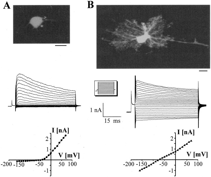
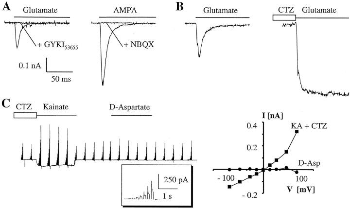
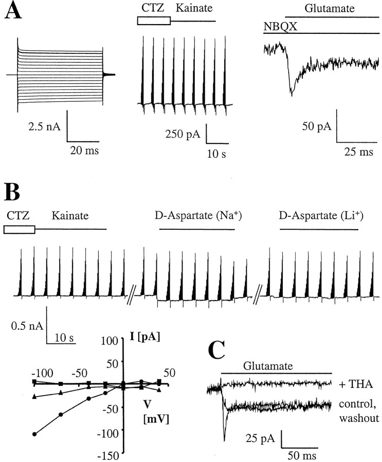
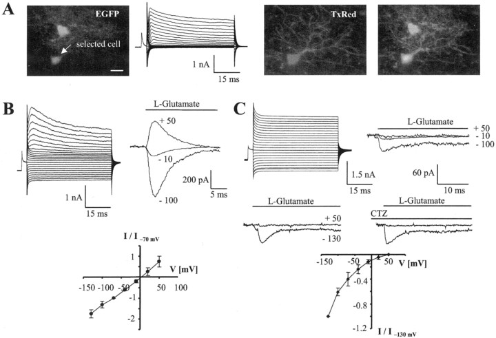
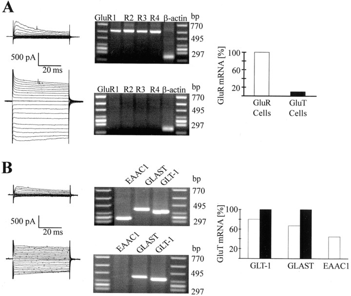
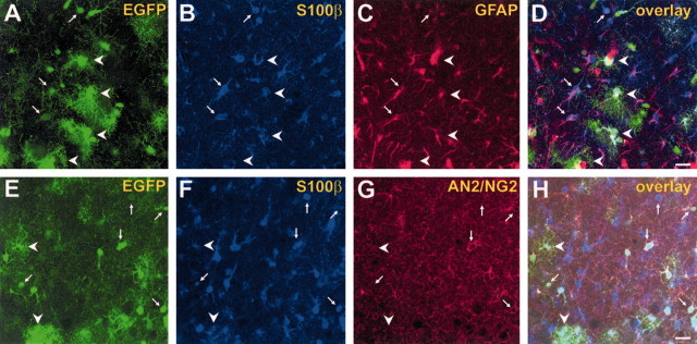
Recent data have suggested the existence of direct signaling pathways between glial cells and neurons. Here we report the coexistence of distinct types of cells expressing astrocyte-specific markers within the hippocampus that display diverse morphological, molecular, and functional profiles. Usage of transgenic mice with GFAP promoter-controlled enhanced green fluorescent protein (EGFP) expression allowed the identification of astroglial cells after fresh isolation or in brain slices. Combining patch-clamp recordings and single-cell reverse transcription-PCR, we distinguished two morphologically distinct types of EGFP-positive cells, one expressing glutamate transporters and the other expressing ionotropic glutamate receptors. None of the EGFP-positive cells coexpressed glutamate receptors and transporters. Subpopulations of glutamate receptor-bearing EGFP-positive cells expressed AN2, the mouse homolog of the rat NG2 proteoglycan or transcripts for excitatory amino acid carrier 1, a neuronal glutamate transporter. Our data demonstrate the presence of distinct, independent populations of cells with astroglial properties in the developing hippocampus that can differently modulate neuronal signaling pathways. The observed heterogeneity of cells with GFAP promoter-regulated EGFP expression and S100beta/GFAP immunoreactivity challenges the hitherto accepted definition of astrocytes.


Figures

Fig. 1.
Fig. 1.
Whole-cell recordings from morphologically distinct EGFP-positive cells acutely isolated from the CA1 stratum radiatum of Tg(GFAP/EGFP) mice. After a prepulse to −110 mV, the membrane was stepped from −160 to +70 mV (10 mV increments; seeinset). A, The current pattern of the weakly fluorescent cell was dominated by outward rectifying K+ currents, whereas only negligible inward currents were activated after hyperpolarization. TheIV relation demonstrates outward rectification of the peak currents (P13; bottom).B, The current phenotype of the brightly fluorescent, widely branched cell differed from the aforementioned cell type in the prominent inward currents. Note the almost linearIV relationship of this P12 EGFP-positive cell (peak currents; bottom). Scale bar, 10 μm.
Fig. 2.
Fig. 2.
Weakly fluorescent acutely isolated cells express functional AMPA receptors but not glutamate transporters.A, In two different cells, rapid application of glutamate (left, P9) or AMPA (right, P15) evoked fast transient and completely desensitizing (τ = 6 and 8.2 msec, respectively) inward currents that were fully inhibited by the AMPA receptor antagonist GYKI53655 and NBQX. B, In another EGFP-positive cell (P13), the control response to glutamate (left) was enhanced twofold when the same cell was exposed to CTZ before application of the agonist (right). C, Membrane currents were elicited in a P13 cell by stepping the membrane between −100 and +100 mV for 100 msec (100 msec intervals) in a bath solution containing K+ channel blockers. The inset gives one current family at higher resolution. The cell displayed GluR currents after application of CTZ and kainate, whereasd-aspartate failed to evoke responses. TheIV relationships (right) were calculated by subtracting current amplitudes at corresponding voltages in the presence of kainate or d-aspartate from the control currents recorded before application of the respective substance.
Fig. 3.
Fig. 3.
Brightly fluorescent, highly branched cells after acute isolation possess functional glutamate transporters but lack AMPA receptors. A, In an EGFP-positive cell at P14, prominent time- and voltage-independent currents were activated (left). Subsequent application of CTZ and kainate failed to evoke a response (for stimulation protocol, see legend to Fig.2C). However, inward currents were elicited in the same cell after fast application of glutamate (1 mm) in the presence of NBQX (right). B, Another example of the absence of coexpression of glutamate receptors and transporters. The cell (P11) did not respond to CTZ and kainate, but inward currents were evoked by d-aspartate, with the latter ceasing when Na+ was replaced with Li+ in the bath solution. The bottom panel gives the respective IVrelationships. Na+, circles; Li+,triangles; kainate/CTZ, squares.C, Fast application of glutamate (0.5 mm, duration 10 sec) to an EGFP-positive cell at P14 activated an inward current displaying an initial, rapidly decaying (τ = 3 msec) and a sustained component. Coapplication of THA led to a complete, partly reversible inhibition of the response.
Fig. 4.
Fig. 4.
Properties of GluR cells and GluT cells in situ. A, Distinct intrinsic fluorescence of a GluT cell (top) and a GluR cell (arrow) in the CA1 stratum radiatum of a P12 mouse (left). Whole-cell currents were obtained from the GluR cell (see Fig. 1 for stimulation protocol, KCl-based pipette solution). During recording, the cell was filled with Texas Red-conjugated dextran, revealing thin, branched processes that were not visible in the EGFP image. Superposition of the fluorescence images demonstrated a close association of both astrocytes (right).B, Current pattern of another GluR cell (P12). Intracellular replacement of KCl with KSCN gave rise to a prominent resting conductance. Subsequently, an outside-out patch was excised from the cell, and glutamate was rapidly applied to the patch at different membrane potentials (top right). To get meanIV relationships, the responses of different cells (n = 10) were normalized to the respective peak amplitudes at −70 mV and averaged (bottom). C, A GluT cell (P18) was investigated as described in B. Note the absence of outward currents after glutamate application to the outside-out patch (top right). Even preapplication of CTZ to the patch failed to disclose any glutamate responses at positive voltages (middle). The IV curve gives mean values obtained from different GluT cells (n = 6) after normalizing to maximum currents at −130 mV (bottom).
Fig. 5.
Fig. 5.
Segregated expression of AMPA receptor and glutamate transporter transcripts by individual acutely isolated EGFP-positive cells. A, Presumed GluR cells and GluT cells were selected under fluorescence illumination, and membrane currents were activated as described in Figure 3A. After recording, the cytoplasm was harvested for RT-PCR. The GluR cell (top) contained mRNAs for all four GluR subunits, whereas only the housekeeping gene was detected in the GluT cell (middle). The bar graph (bottom) gives the frequency of GluR mRNA detection in both cell types (GluR cells,n = 6; GluT cells, n = 10).B, Both cell types were tested for the expression of transporter transcripts. A coexpression of EAAC1, GLAST, and GLT-1 was found in a GluR cell (top). The GluT cell (middle) contained mRNAs encoding GLAST and GLT-1. The bar graph (bottom) summarizes the distribution of transporter RNAs in the two cell types. The GluT cells (n = 17) (filled bars) all coexpressed GLAST and GLT-1, whereas only a partial overlap of both transcripts was observed in the GluR cells (n = 18) (open bars). Some of the GluR cells (4 of 9) also contained mRNA for EAAC1, whereas this transcript was absent in GluT cells (n = 10).
Fig. 6.
Fig. 6.
Morphologically and immunohistochemically distinct populations of EGFP-positive cells can be distinguished in the CA1 stratum radiatum of Tg(GFAP/EGFP) mice (P10).AD, Triple-fluorescence analysis, using intrinsic EGFP fluorescence (green) and double-immunolabeling against S100β (blue) and GFAP (red). Arrows indicate weakly EGFP fluorescent, GluR cells expressing GFAP and S100β.Arrowheads mark S100β- and GFAP-positive astrocytes with high levels of EGFP resembling GluT cells.EH, Triple-fluorescence analysis with intrinsic EGFP fluorescence (green) and double-immunolabeling against S100β (blue) and AN2 (red). Arrows mark putative GluR cells with fewer processes that express EGFP, S100β, and AN2.Arrowheads indicate highly branched, EGFP- and S100β-positive cells lacking AN2 immunoreactivity. Note that the fluorescence intensities of GluR cell somata (arrows) saturate the green CLSM channel. The gain settings were increased to visualize thin processes with low EGFP expression. Scale bar, 20 μm.

References

    1. Akopian G, Kuprijanova E, Kressin K, Steinhäuser C. Analysis of ion channel expression by astrocytes in red nucleus brain stem slices of the rat. Glia. 1997;19:234–246. - PubMed
    1. Anderson CM, Swanson RA. Astrocyte glutamate transport: review of properties, regulation, and physiological functions. Glia. 2000;32:1–14. - PubMed
    1. Araque A, Sanzgiri RP, Parpura V, Haydon PG. Calcium elevation in astrocytes causes an NMDA receptor-dependent increase in the frequency of miniature synaptic currents in cultured hippocampal neurons. J Neurosci. 1998;18:6822–6829. - PMC - PubMed
    1. Arriza JL, Fairman WA, Wadiche JI, Murdoch GH, Kavanaugh MP, Amara SG. Functional comparisons of three glutamate transporter subtypes cloned from human motor cortex. J Neurosci. 1994;14:5559–5569. - PMC - PubMed
    1. Barbour B, Brew H, Attwell D. Electrogenic uptake of glutamate and aspartate into glial cells isolated from the salamander(ambystoma) retina. J Physiol (Lond) 1991;436:169–193. - PMC - PubMed

Publication types

MeSH terms

LinkOut - more resources