. 2003 Mar 1;23(5):1792-803.
doi: 10.1523/JNEUROSCI.23-05-01792.2003.

Leukemia inhibitory factor is a key signal for injury-induced neurogenesis in the adult mouse olfactory epithelium

Affiliations

Leukemia inhibitory factor is a key signal for injury-induced neurogenesis in the adult mouse olfactory epithelium

S Bauer et al. J Neurosci. .

Abstract

The mammalian olfactory epithelium (OE) is composed of primary olfactory sensory neurons (OSNs) that are renewed throughout adulthood by local, restricted neuronal progenitor cells. The molecular signals that control this neurogenesis in vivo are unknown. Using olfactory bulb ablation (OBX) in adult mice to trigger synchronous mitotic stimulation of neuronal progenitors in the OE, we show the in vivo involvement of a cytokine in the cellular events leading to the regeneration of the OE. We find that, of many potential mitogenic signals, only leukemia inhibitory factor (LIF) is induced before the onset of neuronal progenitor proliferation. The rise in LIF mRNA expression peaks at 8 hr after OBX, and in situ RT-PCR and immunocytochemistry indicate that LIF is upregulated, in part, in the injured neurons themselves. This rise in LIF is necessary for injury-induced neurogenesis, as OBX in the LIF knock-out mouse fails to stimulate cell proliferation in the OE. Moreover, delivery of exogenous LIF to the intact adult OE using an adenoviral vector stimulates BrdU labeling in the apical OE. Taken together, these results suggest that injured OSNs release LIF as a stimulus to initiate their own replacement.


Figures

Fig. 1.
Fig. 1.
LIF mRNA is induced and OMP mRNA disappears after OBX. Autoradiograms are shown of typical RT-PCR data obtained from total olfactory organ RNA from one series of adult mice. Eachband corresponds to one animal killed at the delay indicated on top (in hours) after bilateral OBX. Control tubes were included in each experiment, either without retrotranscribed RNA (without cDNA) or with purified RNAs instead of the cDNAs (with RNA). The sequences amplified are indicated on the left: β-actin as internal control, olfactory marker protein (OMP), and leukemia inhibitory factor (LIF).
Fig. 2.
Fig. 2.
Expression kinetics of candidate mitogens and receptors in the olfactory organ of adult mice after OBX. Semiquantitative RT-PCR data are expressed as ratios to β-actin mRNA for each sample, which were averaged as a function of time after lesion (x-axis displays days after OBX; cc marks sham-operated animals). Each point represents the mean ± SEM of two to four animals, and asterisks indicate significant differences (p < 0.05) for OMP, LIF, and LIFR, as assessed with one-way ANOVA and subsequent Bonferroni test (see Materials and Methods). The OMP signal significantly decreases from 56 hr (2 d + 8 hr) until 6 d after OBX. The LIF signal is significantly increased at 8 and 16 hr after OBX, whereas the LIFR signal decreases at 88 hr (3 d + 16 hr). No other significant variation of mRNA expression is detected in this paradigm.
Fig. 3.
Fig. 3.
LIF protein is induced by OBX. Western blots were performed for LIF and β-actin on extracts from olfactory organs sampled at various times (hours, indicated on top) after bilateral OBX. Duplicate samples from two independent mice are shown for each OBX time point. LIF mobility is known to vary according to carbohydrate composition; in this preparation the LIF band is ∼90 kDa. This band only appears after OBX and is obvious at the earliest time point (4 hr after OBX). LIF signal is no longer apparent at 24 hr. The identity of the other bands is not known, but they are apparent without the LIF primary antibody, unlike the 90 kDa band.
Fig. 4.
Fig. 4.
In situ localization of LIF in the olfactory mucosa. A, Using indirect in situ RT-PCR, LIF mRNA expression 8 hr after unilateral OBX is found mainly in the olfactory epithelium (OE).B, Signal density is decreased when the reaction is restricted to reverse transcription by omission of theTaq polymerase. In both cases (A,B), some labeled cells are found in the sustentacular cell (SC) and the basal cell (BC) layers, and in the lamina propria (LP), under the OE (basal lamina is indicated by thedashed line). C, When reverse transcription is omitted, no signal can be detected. D, Perikarya of olfactory sensory neurons (OSN) were identified on adjacent sections using classical in situhybridization for OMP mRNA, which is a specific marker for terminally differentiated OSNs (Margolis, 1982). E,F, LIF protein is localized using immunostaining.E, Transverse section of OE 8 hr after unilateral OBX shows specific labeling localized in olfactory axon bundles (Ax) of ipsilateral septum. F, No significant signal is found in contralateral, uninjured mucosa. Scale bar, 50 μm.
Fig. 5.
Fig. 5.
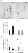
LIF is required for the induction of BrdU labeling in OE after OBX. A, Typical sections of the olfactory organ, through the cartilaginous septum (between thetwo markers at the top of the photographs), are shown stained for BrdU at the peak of proliferation, 5 d after unilateral OBX. When comparing BrdU staining in the septal OE of LIF KO mice and WT littermates, it is clear that proliferation is induced by OBX (OBX,arrows) in the WT OE but not in the LIF KO OE. Cell proliferation in the nonoperated OE (C,arrowheads) is similar for both genotypes. These results were quantified by assessing the number of BrdU-labeled nuclei per linear length of septal OE (arbitrary units) at the peak of proliferation, 5 d after OBX or sham operations, as shown in thegraph (A). The basal level of BrdU labeling indicated by the sham values does not differ between the genotypes. Although BrdU labeling is strongly increased (245% over sham) after OBX in the WT, there is no significant increase in the LIF KO (8% over sham). The difference in BrdU labeling between the genotypes at this time point is highly significant (**p = 0.003). B, The same analysis was performed 2 d after OBX. Interestingly, cell proliferation is significantly decreased compared with sham controls in both the LIF KO mice and WT littermates (**p = 0.003 and *p = 0.04, respectively). Moreover, proliferation is even more reduced in the LIF KO mice (*p = 0.01 when compared with WT value at 2 d). All statistical analysis was performed with Student's t test. Scale bar, 100 μm.
Fig. 6.
Fig. 6.
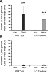
TUNEL labeling in the OE of LIF KO and WT littermates after unilateral OBX. Alternate sections from the same set used for BrdU labeling were used for TUNEL labeling. A, The number of TUNEL-positive nuclei per linear length of OE (arbitrary units) was analyzed at the peak of apoptosis, 2 d after OBX. As expected, there is a large increase in staining in the WT (>500-fold over sham). The increase in TUNEL labeling in the LIF KO OE is also very significant, although somewhat less than in the WT (200-fold). The difference between the genotypes does not reach statistical significance, given the variation and the number of mice available for this experiment (3–5 animals per point). B, At 5 d after OBX, the number of TUNEL-labeled cells declines to nonsignificant values when compared with sham controls in both genotypes (p > 0.05). Statistical analysis was performed with Student's t test (***p < 0.001).
Fig. 7.
Fig. 7.
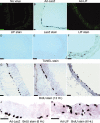
Delivery of LIF using an adenoviral vector induces major increases in TUNEL and BrdU labeling. Normal adult mice were intranasally infected with no virus (A,D, G), with LacZ-expressing virus (B, E, H,J), or with LIF-expressing virus (C, F, I,K). The top row of pictures displays typical OE sections stained with the anti-LIF antibody (A, C) or for β–galactosidase (B). Both viruses infect and cause expression of the transgenes in the OE. The middle row of pictures illustrates results with TUNEL staining. There is a selective increase in TUNEL labeling caused by the LIF vector in all cell layers of the OE (F). Twelve hours after BrdU injection, the LIF vector also promotes the appearance of BrdU labeling specifically in the apical layer of the OE, whereas it decreases labeling in the basal cell layer (I) (see also quantification of staining presented in Table 2). At an earlier time of BrdU labeling (6 hr), infection with the LIF virus yields labeled cells in all layers of the OE (K), and in some cases there appears to be a chain of labeled cells migrating from the basal to the apical layer (K,white asterisk). Scale bar, 30 μm.

References

    1. Banner LR, Patterson PH. Major changes in the expression of the mRNAs for cholinergic differentiation factor/leukemia inhibitory factor and its receptor after injury to adult peripheral nerves and ganglia. Proc Natl Acad Sci USA. 1994;91:7109–7113. - PMC - PubMed
    1. Banner LR, Moayeri NN, Patterson PH. Leukemia inhibitory factor is expressed in astrocytes following cortical brain injury. Exp Neurol. 1997;147:1–9. - PubMed
    1. Bauer S, Mauduit C, Jourdan F, Benahmed M, Moyse E. In vivo involvement of the cytokine LIF during lesion-induced renewal of olfactory sensory neurons in adult mouse. In: Patterson PH, Kordon C, Christen Y, editors. Neuro-immune interactions in neurologic and psychiatric disorders. Springer; Fondation IPSEN. Berlin: 2000. pp. 153–160.
    1. Blesch A, Uy HS, Grill RJ, Cheng JG, Patterson PH, Tuszynski MH. Leukemia inhibitory factor augments neurotrophin expression and corticospinal axon growth after adult CNS injury. J Neurosci. 1999;19:3556–3566. - PMC - PubMed
    1. Buckland ME, Cunningham AM. Alterations in expression of the neurotrophic factors glial cell line-derived neurotrophic factor, ciliary neurotrophic factor and brain-derived neurotrophic factor, in the target-deprived olfactory neuroepithelium. Neuroscience. 1999;90:333–347. - PubMed

Publication types

MeSH terms

LinkOut - more resources