. 2003 Mar 1;23(5):1867-78.
doi: 10.1523/JNEUROSCI.23-05-01867.2003.

Neuronal basis of crossed actions from the reticular formation on feline hindlimb motoneurons

Affiliations

Neuronal basis of crossed actions from the reticular formation on feline hindlimb motoneurons

Elzbieta Jankowska et al. J Neurosci. .

Abstract

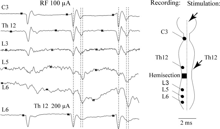
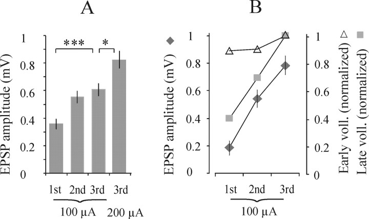
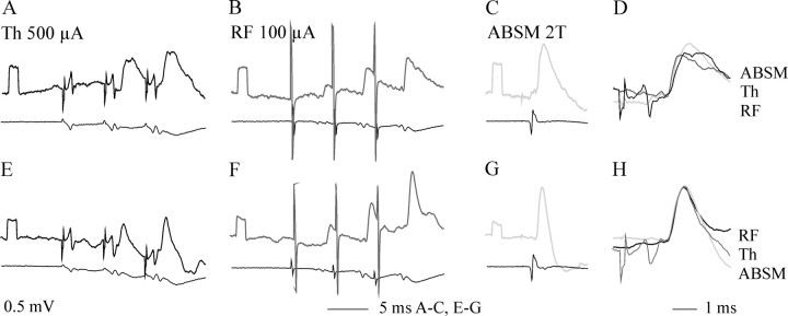
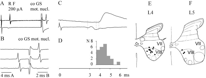
Pathways through which reticulospinal neurons can influence contralateral limb movements were investigated by recording from motoneurons innervating hindlimb muscles. Reticulospinal tract fibers were stimulated within the brainstem or in the lateral funiculus of the thoracic spinal cord contralateral to the motoneurons. Effects evoked by ipsilaterally descending reticulospinal tract fibers were eliminated by a spinal hemisection at an upper lumbar level. Stimuli applied in the brainstem evoked EPSPs, IPSPs, or both at latencies of 1.42 +/- 0.03 and 1.53 +/- 0.04 msec, respectively, from the first components of the descending volleys and with properties indicating a disynaptic linkage, in most contralateral motoneurons: EPSPs in 76% and IPSPs in 26%. EPSPs with characteristics of monosynaptically evoked responses, attributable to direct actions of crossed axon collaterals of reticulospinal fibers, were found in a small proportion of the motoneurons, whether evoked from the brainstem (9%) or from the thoracic cord (12.5%). Commissural neurons, which might mediate the crossed disynaptic actions (i.e., were antidromically activated from contralateral motor nuclei and monosynaptically excited from the ipsilateral reticular formation), were found in Rexed's lamina VIII in the midlumbar segments (L3-L5). The results reveal that although direct actions of reticulospinal fibers are much more potent on ipsilateral motoneurons, interneuronally mediated actions are as potent contralaterally as ipsilaterally, and midlumbar commissural neurons are likely to contribute to them. They indicate a close coupling between the spinal interneuronal systems used by the reticulospinal neurons to coordinate muscle contractions ipsilaterally and contralaterally.


Figures

Fig. 1.
Fig. 1.
Possible substrates of oligosynaptic actions of reticulospinal tract fibers on contralateral hindlimb motoneurons.A, Monosynaptic connections via crossing axon collaterals of RF neurons (1, 2) with cell bodies located either on the opposite or same side as motoneurons and of RF neurons descending at the side of location of motoneurons (3); effects of the latter were eliminated by a hemisection of the spinal cord a few segments rostral to the motoneurons. B, Hypothetical disynaptic connections between reticulospinal tract neurons and contralateral motoneurons via interneurons of the lumbosacral enlargement: commissural neurons (4) and interneurons located at the same side as the motoneurons, the latter activated either by crossing collaterals of the uncrossed reticulospinal tract fibers (5) or by collaterals of the crossed reticulospinal tract fibers (6); effects of the latter were eliminated by a hemisection. C. Alternative disynaptic connections via more rostrally located neurons: long propriospinal tract neurons (7) and other indirectly activated reticulospinal neurons (8) and their crossing segmental axon collaterals. Reticulospinal neurons labeled A,D, and G might represent the same RF neurons. Effects mediated by connections 1,4, and 8 might thus be evoked in parallel. The same may be true for effects mediated by connections2 and 4, if reticulospinal neurons labeled B and F represented the same neurons. Interneurons located on the same side as motoneurons might be excited not only by connections 5 and 6but also by axon collaterals of other reticulospinal neurons and propriospinal neurons; however, these possibilities are not indicated in the diagrams for the sake of simplicity. Any additional synaptic actions of propriospinal neurons or indirectly activated reticulospinal tract neurons mediated by spinal interneurons would, however, be evoked trisynaptically. Int, Interneurons; Com, commissural neurons; PN, long propriospinal neurons;Mn, motoneurons; i, ipsilateral;co, contralateral. Arrows indicate sites of stimulation.
Fig. 2.
Fig. 2.
Temporal facilitation of the second components of descending volleys induced by RF stimuli. Fromtop to bottom are records from the surface of the left lateral funiculus at the indicated levels. All these records are from the same experiment and illustrate descending volleys evoked by stimuli applied in the right MLF and, for comparison, to the right lateral funiculus at T12 (Th12). In thediagram, the stimulation sites are indicated byarrows, and the recording sites are indicated byfilled circles. Amplitudes of the first positive (downward) components at C3 and Th and at L3, L5, and L6 were normalized to aid the comparison of the second components. The records are aligned so that the peaks of the first components of the descending volleys coincide. Shock artifacts are removed, and thick segments of the lines indicate the times of application of the stimuli. The three dashed linescoincide with the peaks of the first components and peaks of the second components at C3 and L6.
Fig. 3.
Fig. 3.
Examples of PSPs evoked in a contralateral GS α motoneuron by RF stimulation. A–C, EPSPs evoked by triple RF stimuli at 60, 70, and 100 μA and corresponding records of descending volleys (top, bottom traces, respectively). D, Extracellular field potentials recorded just outside the motoneuron; note that they displayed a similar temporal facilitation. The arrowindicates the terminal potential. E–G, Effects of single, double, and triple RF stimuli at 100 μA. The gray trace overlying the third EPSP inG is the one from E, normalized to the size of the EPSP evoked the third stimulus. Note the faster decline of the EPSP evoked by the third stimulus, indicating that IPSPs were also evoked. H, Expanded view of the middle part (indicated in F by the dotted horizontal line) of the records in F, with extracellular records of field potentials being subtracted from the intracellularly evoked EPSP to allow a better estimate of EPSP onset. Dotted vertical lines indicate the positive peaks of the first and second components of the descending reticulospinal volley and the onset of the EPSP. Voltage calibration in C is forA–C and E–G. In this and the following figures, the negativity is downward in microelectrode recordings (intracellular and extracellular, usually top traces) and upward in records from the surface of the spinal cord (usually bottom traces).
Fig. 4.
Fig. 4.
Amplitudes of EPSPs evoked from RF.A, Mean amplitudes ± SEM of EPSPs evoked by the first, second, and third stimuli at 100 μA and by the third stimulus at 200 μA. B, Relationships between amplitudes of EPSPs evoked by 100 μA and amplitudes of early and late components of the descending volleys (voll.) evoked by successive stimuli (with respect to those evoked by the third stimulus).B includes data for EPSPs with peak amplitudes exceeding 0.5 mV, which were evoked by the third stimulus in 28 motoneurons in the L7 segment in three experiments (GS, PBST, and FDL). Differences between amplitudes of EPSPs evoked by the third 100 and 200 μA stimuli and between effects of the third and first (but not the second) stimuli are highly statistically significant.
Fig. 5.
Fig. 5.
Examples of EPSPs likely to be evoked monosynaptically in three motoneurons. Top traces are intracellular records from a motoneuron, and bottom traces are from the surface of the spinal cord. Dotted lines indicate the positive peaks of the first component of the descending volley and the onset of the presumably monosynaptically and disynaptically evoked EPSPs. A–C, EPSPs evoked in a DP motoneuron by stimuli applied in the RF (A), at a thoracic level (B), and by group I afferents (C); in C, the last EPSP fromA is superimposed (gray) on the group Ia EPSP after its amplitude has been normalized.D–F, EPSPs evoked in an FDL motoneuron by RF stimuli at two intensities as indicated. Note that the first two weaker stimuli and the first stronger stimulus evoked a short latency EPSP, whereas the following stimuli also evoked a later EPSP. F, First EPSP in D shown expanded. G, H, EPSPs evoked in a PBST motoneuron by stimuli applied at two different depths along the electrode track shown in Figure 10A. Note that EPSPs evoked from the more dorsal site followed each of the three stimuli and appeared at a shorter latency, whereas those evoked from the more ventral site were induced only by the second and third stimuli and at a longer latency (at the level of the third dotted line). I, Extracellular (Extracell.) field potentials evoked by the same stimuli as the records in H: truncated shock artifacts.
Fig. 6.
Fig. 6.
Temporal facilitation of EPSPs evoked from the Th level. A–E, H, Records from two Q motoneurons.A, E, EPSPs evoked by Th stimuli. B, F, EPSPs evoked from the RF. C, G, Monosynaptic EPSPs evoked by group Ia afferents in an ABSM nerve. D, H, Superimposed EPSPs of all three origins, expanded and normalized to the same initial peak amplitude. Note the similar rise time.
Fig. 7.
Fig. 7.
Temporal facilitation of IPSPs.A–D, Examples of IPSPs evoked by RF stimuli in four Q motoneurons (all from the same experiment, along the electrode track illustrated in Fig. 10B). In A–C, the gray traces superimposed on the last PSPs are those evoked by the preceding stimulus to show the faster decline of the last EPSPs, which is an indication that they were followed by IPSPs growing in size after successive stimuli. The superimposed EPSP was normalized in A but at the original size in B andC. D, Records of IPSPs apparently not associated with EPSPs. The superimposed gray trace is that of an IPSP evoked by Q group I afferents in an unidentified flexor motoneuron recorded in the same segment. The amplitude of the latter was normalized to that of the IPSP evoked from the RF to allow a better comparison of their time course. E–H, Records from a Tib motoneuron recorded in another experiment: E, just after penetration of the motoneuron; F, after its depolarization by 20 nA; G, after removal of the polarization current; H, after its hyperpolarization by 10 nA. Note an increase and a much clearer onset of the IPSP (indicated by the dotted line) after the depolarization.
Fig. 8.
Fig. 8.
Lamina VIII commissural neurons antidromically activated from contralateral motor nuclei and monosynaptically excited from RF or MLF. A, Extracellular records from an interneuron (4 superimposed traces), illustrating highly synchronized short-latency responses after stimuli applied in the reticular formation and in the contralateral motor nucleus.B, Single-sweep records from the same interneuron showing that responses from the motor nucleus were prevented from appearing when synaptically evoked spikes preceded them at an interval of approximately twice the peripheral conduction time (the synaptic–antidromic collision test); the shock artifacts are truncated. D, Histogram of times of transmission through the commissural neurons (sums of latencies of the synaptic and antidromic activation) for 24 extracellularly recorded commissural interneurons, which were monosynaptically activated from the reticular formation. The histogram is in the same scale as the record of a disynaptic EPSP evoked in a motoneuron in C (expanded record from Fig. 2H). E, F, Location of 16 intracellularly labeled interneurons of the present sample in the L4 and L5 segments. They were injected with rhodamine dextran and examined under confocal microscopy. The location of these neurons is indicated on diagrams of the gray matter with the borders between Rexed's laminas (Rexed, 1954) indicated.
Fig. 9.
Fig. 9.
Extent of the areas from which disynaptic PSPs were evoked in contralateral motoneurons. A, B, Intracellular records from a GS motoneuron (moton.) showing effects of stimuli (70 μA) applied along the electrode track indicated in Figure 10A, dotted line, and descending (Desc.) volleys evoked by the same stimuli. C, D, Similar series of records from a Sart motoneuron and the descending volleys induced by stimuli (80 μA) applied along the electrode track indicated in Figure10B, dotted line. Double dotted lines in B and D coincide with the first and second components of the descending volleys.
Fig. 10.
Fig. 10.
Stimulation sites in the medulla. A, B, Photomicrographs in the planes of the electrode tracks indicated by the dashed lines from the experiments in which the records illustrated in Figure 9A–D were obtained; the electrolytic lesions were made at H coordinates 6 and 5.5. C, Stimulation sites from which excitation or inhibition of commissural neurons or both were evoked at stimulus intensities ≤100 μA; all these sites were ipsilateral.interneur., Interneurons. D, Stimulation sites from which disynaptic EPSPs, IPSPs, or both were evoked in motoneurons (motoneur.) at stimulus intensities ≤100 μA; those at the left side (ipsilateral) were used to compare effects of stimuli applied at the right side (contralateral). Upward and downward triangles indicate stimulation sites in the same experiment.

Similar articles

Cited by

References

    1. Aggelopoulos NC, Edgley SA. Segmental localisation of the relays mediating crossed inhibition of hindlimb motoneurones from group II afferents in the anaesthetized cat spinal cord. Neurosci Lett. 1995;185:60–64. - PubMed
    1. Aggelopoulos NC, Burton MJ, Clarke RW, Edgley SA. Characterization of a descending system that enables crossed group II inhibitory reflex pathways in the cat spinal cord. J Neurosci. 1996;16:723–729. - PMC - PubMed
    1. Alstermark B, Kummel H. Transneuronal transport of wheat germ agglutinin conjugated horseradish peroxidase into last order spinal interneurones projecting to acromio- and spinodeltoideus motoneurones in the cat. 1. Location of labelled interneurones and influence of synaptic activity on the transneuronal transport. Exp Brain Res. 1990;80:83–95. - PubMed
    1. Burke RE. Composite nature of the monosynaptic excitatory postsynaptic potential. J Neurophysiol. 1967;30:1114–1137. - PubMed
    1. Burke RE. The use of state-dependent modulation of spinal reflexes as a tool to investigate the organization of spinal interneurons. Exp Brain Res. 1999;128:263–277. - PubMed

Publication types

LinkOut - more resources