. 2003 Mar 1;23(5):1924-32.
doi: 10.1523/JNEUROSCI.23-05-01924.2003.

Reversible disorganization of the locomotor pattern after neonatal spinal cord transection in the rat

Affiliations

Reversible disorganization of the locomotor pattern after neonatal spinal cord transection in the rat

Jean-Chrétien Norreel et al. J Neurosci. .

Abstract

The central pattern generators (CPGs) for locomotion, located in the lumbar spinal cord, are functional at birth in the rat. Their maturation occurs during the last few days preceding birth, a period during which the first projections from the brainstem start to reach the lumbar enlargement of the spinal cord. The goal of the present study was to investigate the effect of suppressing inputs from supraspinal structures on the CPGs, shortly after their formation. The spinal cord was transected at the thoracic level at birth [postnatal day 0 (P0)]. We examined during the first postnatal week the capacity of the CPGs to produce rhythmic motor activity in two complementary experimental conditions. Left and right ankle extensor muscles were recorded in vivo during airstepping, and lumbar ventral roots were recorded in vitro during pharmacologically evoked fictive locomotion. Mechanical stimulation of the tail elicited long-lasting sequences of airstepping in the spinal neonates and only a few steps in sham-operated rats. In vitro experiments made simultaneously on spinal and sham animals confirmed the increased excitability of the CPGs after spinalization. A left-right alternating locomotor pattern was observed at P1-P3. Both types of experiments showed that the pattern was disorganized at P6-P7, and that the left-right alternation was lost. Alternation was restored after the activation of serotonergic 5-HT(2) receptors in vivo. These results suggest that descending pathways, in particular serotonergic projections, control the strength of reciprocal inhibition and therefore shape the locomotor pattern in the neonatal rat.


Figures

Fig. 1.
Fig. 1.
Tail stimulation triggers long sequences of airstepping in cord-transected neonates. A, Experimental device adapted from Fady et al. (1998). Animals were supported by means of an adjustable sling. The tail was pinched with a forceps.B, Rectified EMG activities from left and right ankle extensor muscles during airstepping induced by tail pinch (horizontal bar) in a 2-d-old rat that had been spinalized at birth. Bottom traces are shown at an extended time base to illustrate the pattern, consisting of left–right alternation. C, Evolution of the period and burst proportion (burst duration/period) in the left ankle extensor muscles during the episode of airstepping illustrated inB.
Fig. 2.
Fig. 2.
The excitability of CPGs is increased in neonates spinalized at birth. A, In vitroexperiments on spinal cord preparations (T9–S4) isolated from two 2-d-old rats issued from the same litter. One animal had been spinalized at birth (spinal), and the other one had been operated in the same way except for the spinal cord transection (sham). B, C, Rectified ventral root (VR) activities from the spinal (B) and the sham (C) animals during fictive locomotion induced by NMA. Locomotor-like activity was characterized by alternation both between right and left ventral root activities and between L3 and L5 bursting on the same side. Dashed lines indicate the approximate peak of bursts occurring in the right L3 ventral root. D, Relationship between the latency of ventral root bursting (relative to the onset of NMA perfusion) and the NMA concentration in spinal (○) and sham (▪) animals.
Fig. 3.
Fig. 3.
The left–right alternating pattern of airstepping is lost at the end of the first postnatal week in the absence of supraspinal inputs. A1, B1, Rectified EMG activity from left and right ankle extensor muscles in 3-d-old (A1) and 6-d-old (B1) spinal rats. Traces were selected 3–13 sec after the onset of the airstepping episode. Dashed lines indicate the approximate peak of bursts occurring in the right ankle extensor muscles.A2, B2, Cross-correlograms between the left and right EMG recordings illustrating the out-of-phase relationship at P3 (A1) and a synchronization at P6 (B1) between the bursts recorded in the two extensor muscles. Cross-correlation analysis was computed from 20 sec of airstepping activity.C1, C2, Circular histograms showing the distribution of phase relationships between left and right motor bursts at P1–P3 (C1) and P6–P7 (C2). Bars indicate the number of observations within each class range (width, 10°). Data from seven animals in each age group were pooled; ∼40 steps in two episodes were selected for each animal. The mean vector angle was 174.7 ± 5.1° (n = 294) at P1–P3 and 20.9 ± 10.1° (n = 289) at P6–P7; the length of the mean vector was 0.44 and 0.23, respectively. The 99% confidence interval is illustrated.
Fig. 4.
Fig. 4.
The degree of coactivation of left and right ankle extensor muscles during airstepping increased with age in spinal rats.A, Mean correlation coefficient between left and right EMG activities during 10–20 sec episodes of airstepping (40 episodes in 8 animals at P1–P3 and 71 episodes in 12 rats at P6–P7). ***p < 0.001; t test.B, Period of airstepping activity and burst proportion (burst duration/period) in the two age groups. Approximately 20 steps were considered (from the 5th to the 25th step) for analysis in each airstepping episode (314 steps taken into account). ns, Not significant; p > 0.05; Mann–Whitney test. ***p < 0.001; t test.
Fig. 5.
Fig. 5.
The in vitro NMA-induced locomotor-like rhythm is disorganized in spinal rats at P4–P6.A1, Ventral root (VR) activities induced by NMA in a spinal rat at P5.A2, Cross-correlogram between left and right L5 ventral root signals. The analysis was computed from 2 min of a locomotor-like activity similar to that illustrated inA1. B, Mean correlation coefficient between left and right ventral root activities. Spinals, Twenty-nine applications of NMA in 10 experiments; shams, Ten applications on four spinal cords. ***p < 0.001; Mann–Whitney test.
Fig. 6.
Fig. 6.
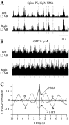
5-HT in vitro switches the NMA-induced disorganized rhythm toward a left–right alternating pattern in spinal rats. A, B, Rectified ventral root (VR) recordings at P6 in the presence of NMA alone (A) or together with 5-HT (B). Dashed lines indicate the approximate peak of bursts occurring in the left L3 ventral root.C, Cross-correlograms between left and right L3 ventral root signals computed from 2 min of activity induced by NMA (solid line) or NMA plus 5-HT (dotted line). Arrowheads point to the successive peaks in the correlogram.
Fig. 7.
Fig. 7.
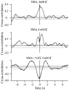
Increasing the excitability of the network does not mimic the effect of 5-HT. Cross-correlograms between left and right L3 ventral root signals computed from 2 min of activity induced by NMA before (top) and after (middle) increasing the extracellular concentration of potassium from 4 to 6 mm, or NMA plus 5-HT (bottom) are shown.
Fig. 8.
Fig. 8.
The left–right alternating pattern reappearsin vivo after the activation of 5-HT2receptors.A1–A4, Cross-correlograms between left and right ankle extensor EMG signals during airstepping, before (A1) and at different times (A3, A4) after the injection of DOI. The analysis was run on 10 sec of airstepping activity, starting 1 sec after the episode onset. B1, 2, Distribution of phase relationships between left and right motor bursts before (B1) and after (B2) the injection of DOI. Same experiment as in A. Mean vector angle: 98.4 ± 9.5°, n = 107 (length, 0.4) before DOI; 181.7 ± 4.1°, n = 153 (length, 0.68) after DOI. The 99% confidence interval is represented. C, Mean correlation coefficient at P1–P3, P6–P7 (identical to Fig.4A), and P6–P7 after DOI injection (48 episodes analyzed in 6 animals). *p < 0.05; **p < 0.01; ***p < 0.001; one-way ANOVA with Tukey post-test.

Similar articles

Cited by

References

    1. Aggelopoulos NC, Burton MJ, Clarke RW, Edgley SA. Characterization of a descending system that enables crossed group II inhibitory reflex pathways in the cat spinal cord. J Neurosci. 1996;16:723–729. - PMC - PubMed
    1. Andén N-E, Häggendal J, Magnusson T, Rosengren E. The time course of the disappearance of noradrenaline and 5-hydroxytryptamine in the spinal cord after transection. Acta Physiol Scand. 1964;62:115–118. - PubMed
    1. Barbeau H, Rossignol S. Recovery of locomotion after chronic spinalization in the adult cat. Brain Res. 1987;412:84–95. - PubMed
    1. Barbeau H, Rossignol S. The effects of serotonergic drugs on the locomotor pattern and on cutaneous reflexes of the adult chronic spinal cat. Brain Res. 1990;514:55–67. - PubMed
    1. Bradley NS, Smith JL. Neuromuscular patterns of stereotypic hindlimb behaviors in the first two postnatal months. II. Stepping in spinal kittens. Brain Res. 1988;466:53–67. - PubMed

Publication types

MeSH terms