

One-trial memory for object-place associations after separate lesions of hippocampus and posterior parahippocampal region in the monkey
- PMID: 12629201
- PMCID: PMC6741967
- DOI: 10.1523/JNEUROSCI.23-05-01956.2003
One-trial memory for object-place associations after separate lesions of hippocampus and posterior parahippocampal region in the monkey
Abstract
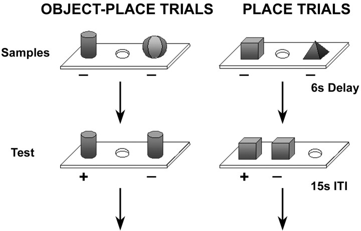
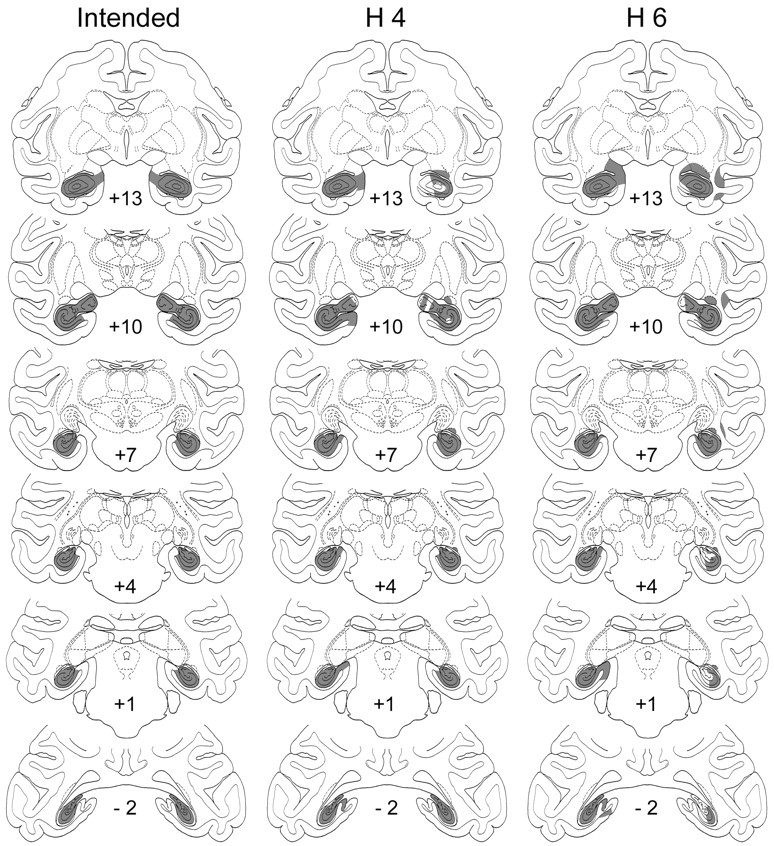
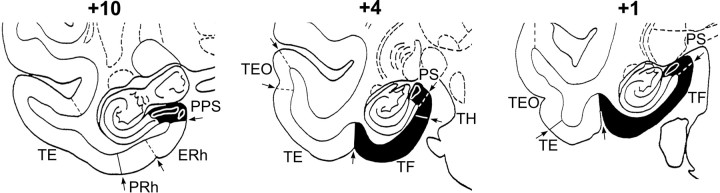
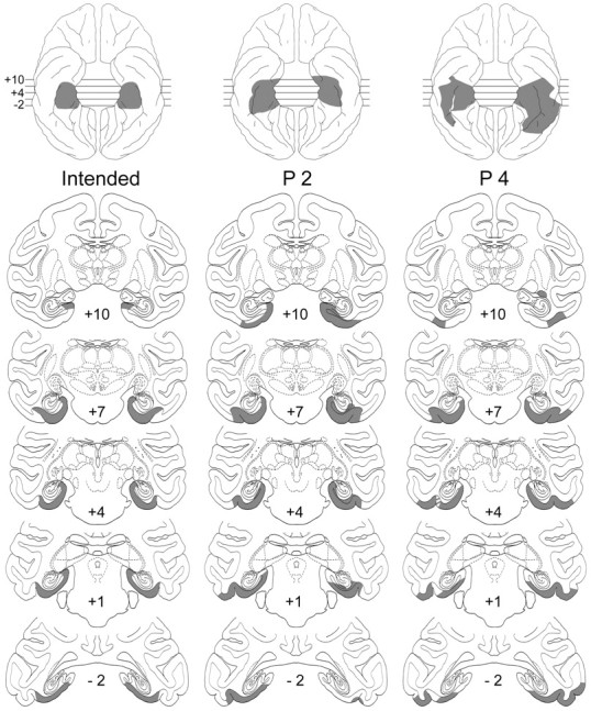
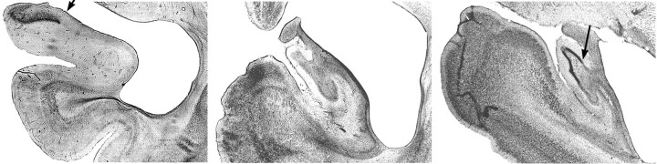
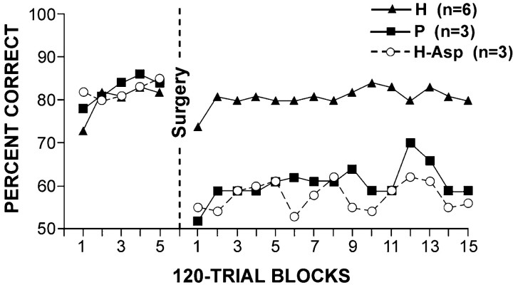
In earlier studies of one-trial spatial memory in monkeys (Parkinson et al., 1988; Angeli et al., 1993), severe and chronic memory impairment for both object-place association and place alone was found after ablation of the hippocampal formation. The results appeared to provide the first clear-cut evidence in the monkey of the essential role of the hippocampus in spatial memory, but that interpretation neglected the inclusion in the lesion of the underlying posterior parahippocampal region. To determine the separate contributions of the hippocampus and posterior parahippocampal region to these spatial forms of one-trial memory, we trained 10 rhesus monkeys, as before, to remember the spatial positions of either two different trial-unique objects overlying two of the wells in a three-well test tray (object-place trials) or simply two of the three wells (place trials). Six of the monkeys then received ibotenic acid lesions restricted to the hippocampal formation (group H), and the four others received selective ablations of the posterior parahippocampal region (group P), comprising mainly parahippocampal cortex, parasubiculum, and presubiculum. Group H was found to be completely unaffected postoperatively on both types of trials, whereas group P sustained an impairment on both types equal in magnitude to that observed after the combined lesions in the original studies. Thus, contrary to the previous interpretation, one-trial memory for object-place association and, perhaps more fundamentally, one-trial memory for two different places appear to be critically dependent not on the hippocampal formation but rather on the posterior parahippocampal region.
Figures
References
-
- Adlam A, Incisa della Rocchetta A, Gadian D, Mishkin M, Vargha-Khadem F. Recognition memory in patients with developmental amnesia. Soc Neurosci Abstr. 2002;28:582.10.
-
- Aguirre GK, Detre JA, Aslop DC, D'Esposito M. The parahippocampus subserves topographical learning in man. Cereb Cortex. 1996;6:823–829. - PubMed
-
- Alvarado MC, Wright AA, Bachevalier J. Object and spatial relational memory in adult rhesus monkeys is impaired by neonatal lesions of the hippocampal formation but not the amygdaloid complex. Hippocampus. 2002;12:421–433. - PubMed
-
- Andersen RA, Asanuma C, Essick G, Siegel RM. Corticocortical connections of anatomically and physiologically defined subdivisions within the inferior parietal lobule. J Comp Neurol. 1990;296:65–113. - PubMed
Publication types
MeSH terms
Substances
Grants and funding
LinkOut - more resources
Full Text Sources
Medical