. 2003 Mar 18;100(6):3239-44.
doi: 10.1073/pnas.0736401100. Epub 2003 Mar 10.

The topoisomerase IIbeta circular clamp arrests transcription and signals a 26S proteasome pathway

Affiliations

The topoisomerase IIbeta circular clamp arrests transcription and signals a 26S proteasome pathway

Hai Xiao et al. Proc Natl Acad Sci U S A. .

Abstract

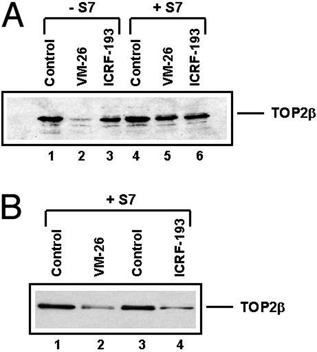
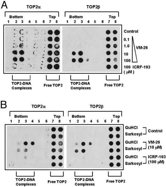
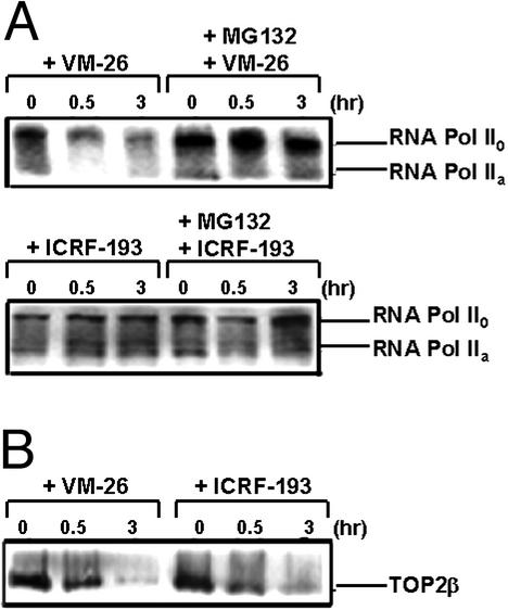
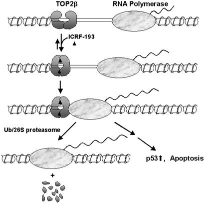
It has been proposed that the topoisomerase II (TOP2)beta-DNA covalent complex arrests transcription and triggers 26S proteasome-mediated degradation of TOP2beta. It is unclear whether the initial trigger for proteasomal degradation is due to DNA damage or transcriptional arrest. In the current study we show that the TOP2 catalytic inhibitor 4,4-(2,3-butanediyl)-bis(2,6-piperazinedione) (ICRF-193), which traps TOP2 into a circular clamp rather than the TOP2-DNA covalent complex, can also arrest transcription. Arrest of transcription, which is TOP2beta-dependent, is accompanied by proteasomal degradation of TOP2beta. Different from TOP2 poisons and other DNA-damaging agents, ICRF-193 did not induce proteasomal degradation of the large subunit of RNA polymerase II. These results suggest that proteasomal degradation of TOP2beta induced by the TOP2-DNA covalent complex or the TOP2 circular clamp is due to transcriptional arrest but not DNA damage. By contrast, degradation of the large subunit of RNA polymerase II is due to a DNA-damage signal.


Figures

Figure 1
Figure 1
ICRF-193 induces time-dependent reduction in the hTOP2β protein level in mammalian cells. HeLa (A) and ZR75-1 (B) cells were treated with 100 μM ICRF-193 for different times (0, 2, 4, and 6 h). Cells then were lysed with the alkaline lysis procedure with staphylococcal S7 nuclease treatment as described in Materials and Methods. Cell lysates were analyzed by immunoblotting with anti-hTOP2α, anti-hTOP2β, and anti-hTOP1 antibodies, respectively. (C) HL-60 cells were treated with VM-26 (25 and 50 μM) in the presence or absence of ICRF-193 (100 μM) for 30 min. Cells then were lysed with the alkaline lysis solution without staphylococcal S7 nuclease treatment. Cell lysates were analyzed by immunoblotting with anti-hTOP2α and anti-hTOP2β antibodies, respectively.
Figure 2
Figure 2
Effect of caspase inhibitor and metabolic inhibitors on ICRF-193-induced down-regulation of hTOP2β. (A) HL-60 cells were treated with ICRF-193 (100 μM) in the presence or absence of the caspase inhibitor Z-Asp(OCH3)-Glu(OCH3)-Val-Asp(OCH3)-fluoromethyl ketone (Z-DEVD-FMK, 50 μM) for 4 h. (B) HL-60 cells were treated with ICRF-193 (0, 1, 10, and 100 μM, lanes 1–4, respectively), MG132 (10 μM, lane 5), and MG132 (10 μM) plus ICRF-193 (100 μM, lane 6) for 4 h. (C) HL-60 cells were treated with 100 μM ICRF-193 for 4 h in the presence and absence of 150 μM DRB or 25 μM CPT. (D) HL-60 cells were treated with 100 μM ICRF-193 for 4 h in the presence or absence of 50 μM cycloheximide (CHX) or 10 μM aphidicolin (APH). Cells were lysed by the alkaline lysis method, and cell lysates were analyzed by immunoblotting with anti-hTOP2α and anti-hTOP2β antibodies, respectively.
Figure 3
Figure 3
ICRF-193 inhibits transcription. Transcription was monitored by 3H-labeled uridine incorporation. HL-60 and HL-60/MX2 cells (A) or mouse embryo fibroblast TOP2β(+/+) and TOP2β(−/−) cells (B) were treated with VM-26 (25 μM), ICRF-193 (200 μM), CPT (25 μM), or DRB (200 μM) for 30 min followed by a 15-min incubation with 3H-labeled uridine. The amount of uridine incorporated into RNA was determined by counting in a scintillation counter. The percentage of transcription inhibition was normalized to cells without drug treatment.
Figure 4
Figure 4
ICRF-193 up-regulates p53 and induces TOP2-dependent apoptosis. (A) The breast cancer ZR75-1 cells were treated with demethylepipodophyllotoxin ethylidene-β-d-glucoside (25 μM), CPT (25 μM), or ICRF-193 (100 μM) for 1 h. Treated cells were lysed directly with SDS sample buffer and prepared for immunoblotting with anti-p53 and anti-actin antibodies, respectively. (B) HL-60 cells and HL-60/MX2 cells (TOP2-deficient mutant cells) were treated with VM-26 (2.5 μM), CPT (2.5 μM), DRB (100 μM), or increasing concentrations of ICRF-193 (25, 100, and 400 μM) for 4 h. Cells then were lysed and processed for detection of the nucleosomal DNA ladders.
Figure 5
Figure 5
ICRF-193 does not induce TOP2–DNA covalent cleavable complexes in HL-60 cells. (A) HL-60 cells were treated with ICRF-193 (200 μM) or VM-26 (100 μM) for 30 min and lysed with the alkaline lysis procedure as described in Materials and Methods. Cell lysates were treated with (+S7) or without (−S7) staphylococcal nuclease S7. Treatment with the nuclease releases hTOP2β from covalent TOP2–DNA complexes, which migrate more slowly (not shown in the gel) than free hTOP2β. (B) Cells were treated with ICRF-193 (100 μM) or VM-26 (100 μM) for 4 h. Cells were lysed and processed for immunoblotting with anti-hTOPβ antibodies as described in the Fig. 2 legend.
Figure 6
Figure 6
ICRF-193 does not induce detectable amounts of covalent TOP2–DNA complexes as determined by the ICE assay. (A) HL-60 cells were treated with ICRF-193 (100 μM) and different concentrations of VM-26 for 30 min. Cells were collected and lysed with 1% Sarkosyl and loaded onto a preformed CsCl step gradient. Centrifugation and immunoblotting of the fractions were performed as described in Materials and Methods. Covalent TOP2–DNA complexes sedimented near the bottom of the gradient (see fractions marked “TOP2–DNA complexes”), whereas free TOP2 enzymes sedimented near the top of the gradient (see fractions marked “Free TOP2”). (B) HL-60 cells were treated with ICRF-193 (100 μM) and VM-26 (100 μM) for 30 min and lysed with either 1% Sarkosyl or 6 M Gdn⋅HCl (GuHCl). The ICE assay was performed as described for A.
Figure 7
Figure 7
VM-26 but not ICRF-193 induces degradation of Pol II. Breast cancer ZR75-1 cells (106 cells per sample) were treated with ICRF-193 (100 μM) or VM-26 (100 μM) for 30 min and 3 h in the presence or absence of the 26S proteasome inhibitor MG132 (1 μM). Cells were then lysed with the alkaline lysis procedure and immunoblotted as described in Materials and Methods. (A) Degradation of the large subunit of Pol II. Pol IIa and Pol II0 were detected by immunoblotting with ARNA-3 antibodies. (B) Degradation of TOP2β. Cell lysates were analyzed by immunoblotting with anti-TOP2β antibodies.
Figure 8
Figure 8
A proposed model for ICRF-193-induced degradation of TOP2β. In the presence of ICRF-193 and ATP, TOP2β is trapped as a closed circular clamp on DNA. This clamp blocks the movement of the transcription-elongation complex. Arrest of transcription triggers 26S proteasome-dependent degradation of TOP2β.

References

    1. D'Arpa P, Liu L F. Biochim Biophys Acta. 1989;989:163–177. - PubMed
    1. Li T K, Liu L F. Annu Rev Pharmacol Toxicol. 2001;41:53–77. - PubMed
    1. Kaufmann S H. Biochim Biophys Acta. 1998;1400:195–211. - PubMed
    1. Mao Y, Desai S D, Ting C Y, Hwang J, Liu L F. J Biol Chem. 2001;276:40652–40658. - PubMed
    1. Desai S D, Li T K, Rodriguez-Bauman A, Rubin E H, Liu L F. Cancer Res. 2001;61:5926–5932. - PubMed

Publication types

MeSH terms

LinkOut - more resources