. 2003 Jul;285(1):F40-8.
doi: 10.1152/ajprenal.00404.2002. Epub 2003 Mar 11.

Urinary excretion of viable podocytes in health and renal disease

Affiliations

Urinary excretion of viable podocytes in health and renal disease

Stefanie U Vogelmann et al. Am J Physiol Renal Physiol. 2003 Jul.

Abstract

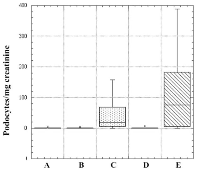
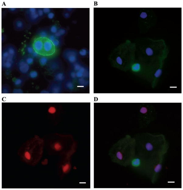
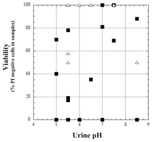
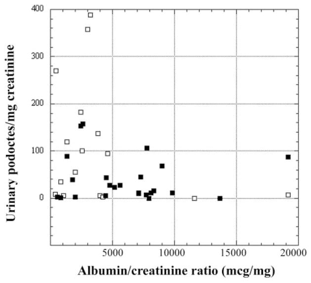
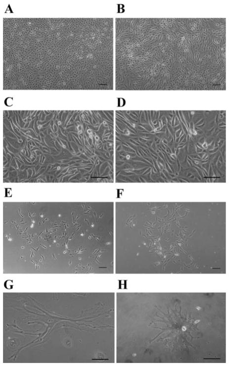
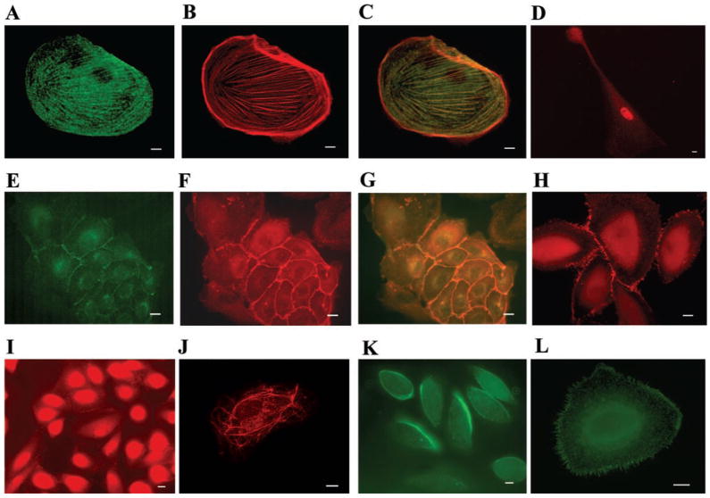
The loss of glomerular visceral epithelial cells (podocytes) has been associated with the development of glomerular sclerosis and loss of renal function. Viability of podocytes recovered from urine of subjects with glomerular disease and of healthy controls was investigated by propidium iodide exclusion and TUNEL staining. Podocyte loss was quantified by cytospin. The growth behavior in culture of urinary cells and their expression of specific markers were examined. The majority of urinary podocytes are viable, although apoptosis occurs in about one-half of the cells. Patients with active glomerular disease excreted up to 388 podocytes/mg creatinine, whereas healthy controls and patients with quiescent disease generally excreted <0.5 podocytes/mg creatinine. The identity of cultured cells was confirmed by their morphology, growth behavior, and expression of podocyte-specific markers. The difference in growth behavior between healthy controls and subjects with active glomerular disease suggests that in active disease viable podocytes detach from the glomerular tuft due to local environmental factors rather than defects in the podocytes per se, whereas in healthy individuals mostly senescent podocytes are shed.


Figures

Fig. 1
Fig. 1
Podocyte excretion expressed as podocytes/mg creatinine. Comparison between urine specimens from healthy individuals and patients with glomerular disease is shown. Five groups are expressed as box plots: A, control subjects; B, inactive focal segmental glomerulosclerosis (FSGS); C, active FSGS; D, inactive lupus nephritis; E, active lupus nephritis. Median values (podocytes/mg creatinine) for the groups (horizontal line within box): A, 0; B, 0.47; C, 19; D, 0.17; E, 74.8.
Fig. 2
Fig. 2
Urine sediment cytospun onto polylysine-coated slide and stained with antipodocalyxin antibody, Hoechst nuclear stain, and propidium iodide. A: podocalyxin-positive cell (binucleate) with podocalyxin in green and nuclear stain in blue. B: podocalyxin-positive cell (green), surrounded by podocalyxin-negative cells (visible by Hoechst nuclear stain in blue). C: propidium iodide (red) stains nuclei of the podocalyxin-negative cells only, indicating lack of viability. D: merged image with anti-podocalyxin staining. Scale bar 10 μm.
Fig. 3
Fig. 3
Podocyte viability [percentage of podocalyxin-positive cells that were propidium iodide (PI) negative] as a function of urinary pH in healthy controls (△) and patients with glomerular disease (■). Viability is not influenced by the pH of the urine.
Fig. 4
Fig. 4
TdT-mediated dUTP-nick-end labeling (TUNEL) staining of podocalyxin-positive cells in sediment confirmed the presence of apoptotic podocytes in urine of both patients and controls. Three podocytes, identified on cytospin with anti-podocalyxin antibody. Two of them are nonapoptotic and one is apoptotic. Antipodocalyxin staining is in green, nuclear stain is in blue, and TUNEL staining is in red. A: urine sediment stained with anti-podocalyxin antibody and Hoechst nuclear stain. B: TUNEL staining with 1 TUNEL-positive cell. C: merged image. Scale bar 10 μm.
Fig. 5
Fig. 5
Relationship of podocyturia to albuminuria in patients with active FSGS (■) and lupus nephritis (□). Active disease is defined as presence of an albumin-to-creatinine ratio >300 μg/mg. Notice the absence of a simple positive relationship of podocyturia to albuminuria.
Fig. 6
Fig. 6
A, C, E: cells cultured from sediments of patient urine. B, D, F: podocytes grown out of explanted glomeruli isolated from healthy parts of a kidney removed for localized tumor. A and B: early dense, cobblestone-like pattern. C and D: cultured cells undergoing crisis. E and F: differentiated podocytes. Comparison between cultured sediments and outgrowths shows substantial similarity. G and H: urinary podocytes with primary and secondary processes. Scale bar 100 μm.
Fig. 7
Fig. 7
Immunofluorescence of cultured urinary cells grown on collagen-coated coverslips. A: anti-synaptopodin staining (green). B: phalloidin staining (red). C: merged image shows colocalization of synaptopodin and actin. D: podocyte expressing WT1 (red). E: cultured cells expressing P-cadherin at cell-cell contact sites (green). F: cultured, cobblestone-like cells expressing ZO-1 at cell-cell contact sites (red). G: merged image. H: more differentiated podocytes expressing ZO-1 at rudimentary slit diaphragms (red). I: podocin staining in less differentiated cells at cell-cell contact sites (red). J: podocin staining in a filamentous pattern (red). K: podocalyxin staining the apical membrane of cells early in culture (green). L: antipodocalyxin staining of filopodia later in culture (green). Scale bar 10 μm.

References

    1. Detrisac CJ, Mayfield RK, Cowell JA, Garvin AJ, Sens DA. In vitro culture of cells exfoliated in the urine by patients with diabetes mellitus. J Clin Invest. 1983;71:170–173. - PMC - PubMed
    1. Drumond MC, Deen WM. Structural determinants of glomerular hydraulic permeability. Am J Physiol Renal Fluid Electrolyte Physiol. 1994;266:F1–F12. - PubMed
    1. Endlich K, Kriz W, Witzgall R. Update in podocyte biology. Curr Opin Nephrol Hypertens. 2001;10:331–340. - PubMed
    1. Fries JW, Sandstrom DJ, Meyer TW, Rennke HG. Glomerular hypertrophy and epithelial cell injury modulate progressive glomerulosclerosis in the rat. Lab Invest. 1989;60:205–218. - PubMed
    1. Hara M, Yamamoto T, Yanagihara T, Takada T, Itoh M, Adachi Y, Yoshizumi A, Kawasaki K, Kihara I. Urinary excretion of podocalyxin indicates glomerular epithelial cell injuries in glomerulonephritis. Nephron. 1995;69:397–403. - PubMed

Publication types

MeSH terms

LinkOut - more resources