. 2003 Mar;14(3):858-70.
doi: 10.1091/mbc.e02-08-0532.

Grb2 regulates internalization of EGF receptors through clathrin-coated pits

Affiliations

Grb2 regulates internalization of EGF receptors through clathrin-coated pits

Xuejun Jiang et al. Mol Biol Cell. 2003 Mar.

Abstract

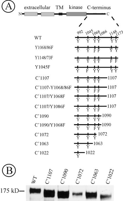
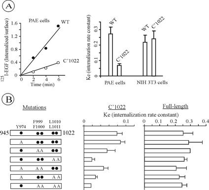
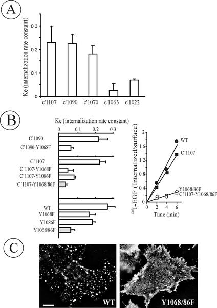
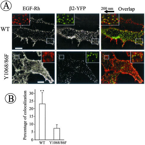
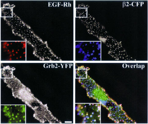
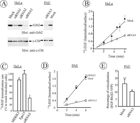
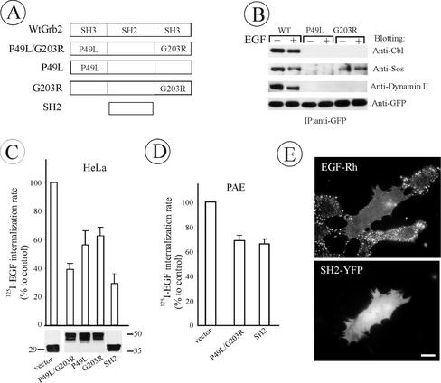
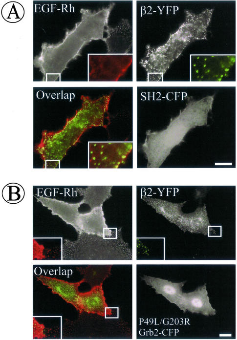
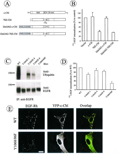
The molecular mechanisms of clathrin-dependent internalization of epidermal growth factor receptor (EGFR) are not well understood and, in particular, the sequence motifs that mediate EGFR interactions with coated pits have not been mapped. We generated a panel of EGFR mutants and stably expressed these mutants in porcine aortic endothelial (PAE) cells. Interestingly, mutations of tyrosine phosphorylation sites 1068 and 1086 that interact with growth-factor-receptor-binding protein Grb2 completely abolished receptor internalization in PAE cells. Quantitative analysis of colocalization of EGF-rhodamine conjugate and coated pits labeled with yellow-fluorescent-protein-tagged beta2 subunit of clathrin adaptor complex AP-2 revealed that EGFR mutants lacking Grb2 binding sites do not efficiently enter coated pits. The depletion of Grb2 from PAE as well as HeLa cells expressing endogenous EGFRs by RNA interference substantially reduced the rate of EGFR internalization through clathrin-dependent pathway, thus providing the direct evidence for the important role of Grb2 in this process. Overexpression of Grb2 mutants, in which the SH3 domains were either deleted or inactivated by point mutations, significantly inhibited EGFR internalization in both PAE and HeLa cells. These findings indicate that Grb2, in addition to its key function in signaling through Ras, has a major regulatory role at the initial steps of EGFR internalization through clathrin-coated pits. Furthermore, the EGFR mutant lacking Grb2 binding sites did not efficiently recruit c-Cbl and was not polyubiquitinated. The data are consistent with the model whereby Grb2 participates in EGFR internalization through the recruitment of Cbl to the receptor, thus allowing proper ubiquitylation of EGFR and/or associated proteins at the plasma membrane.


Figures

Figure 1
Figure 1
Schematic representation of EGF receptor mutants. (A) Wild-type EGFR (WT) is drawn with the extracellular, transmembrane (TM) and intracellular domain (kinase and C terminus). Main tyrosine phosphorylation sites (residues 992, 1068, 1086, 1148, and 1173) and a Cbl binding site (Tyr1045) are indicated in WT receptor. Mutants have C-terminal truncation of 79 (C′1107), 96 (C′1090), 114 (C′1072), 123 (C′1063), and 164 amino acid residues (C′1022). Tyrosine substitutions by phenylalanines (F) are indicated. (B) To confirm the correct size of truncated EGFR mutants, PAE cells expressing WT, C′1107, C′1090, C′1072, C′1063, or C′1022 were lysed, and the EGFR was detected in lysates by immunoblotting with antibody 2913.
Figure 2
Figure 2
125I-EGF Internalization by C′1022 truncated EGFR mutants. (A) The internalization of 1 ng/ml 125I-EGF was measured in PAE cells expressing wild-type (WT) or C′1022 mutant. The rate of internalization is expressed as ratio of internalized and surface 125I-EGF at each time point (left). The internalization rate constant (ke) values measured using 1 ng/ml 125I-EGF in PAE cells were compared with ke values measured under the same conditions (as shown on the left) in NIH 3T3 expressing wild-type or Dc165 mutant EGFR (corresponding to C′1021 truncation; Alvarez et al., 1995; right). The averaged values from multiple experiments with three independent clones of C′1022 PAE cells are presented. Error bars, SEs. (B) Schematic representation of single, double, triple, or quadruple point mutants of C′1022 or full-length EGFR (residues 945-1022 are shown). Tyrosine (Y974), phenylalanines (F999/1000), and leucines (L1010/1011) were substituted by alanines (A, left). The internalization rate constant (ke) of the site-point mutants of C′1022 (panel) and full-length (panel) EGFR were measured using 1 ng/ml 125I-EGF as in A. The bars represent averaged values from multiple experiments with 2–4 independent cell clones for each mutants and the error bars represent SEs.
Figure 3
Figure 3
Tyrosine 1068 and 1086 are critical for EGFR internalization. (A) The ke values were measured in PAE cells stably expressing C′1107, C′1090, C′1072, C′1063, or C′1022 truncated EGFRs as in Figure 2. (B) ke values were measured in cells expressing single or double Tyr1068/1086 mutants of full-length or truncated EGFRs (left) as in A. The bars in A and B represent averaged values from multiple experiments with 2–4 independent cell clones of each mutant and the error bars represent SEs. A representative 125I-EGF internalization experiment performed as in Figure 2A in cells expressing wild-type EGFR (WT), C′1107, C′1107-Y1068/86F, and Y1068/86F mutants is shown on the right. (C) WT and Y1068/86F-expressing PAE cells were incubated with 1 ng/ml EGF-Rh at 37°C for 6 min, and the rhodamine fluorescence images were acquired from living cells. Bar, 10 μm.
Figure 4
Figure 4
Coated pit recruitment of wild-type but not Y1068/86F EGFR. (A) β2-YFP was transiently expressed in wild-type EGFR (WT) and Y1068/86F-expressing PAE cells. The cells grown on coverslips were incubated with 2 ng/ml EGF-Rh at 4°C for 1 h. Cells were then fixed, and the images were acquired through Cy3 (red) and FITC (green) filter channels. Insets: High magnification images of the small regions of the cell shown by white rectangles. In inset overlaps, rhodamine images were shifted approximately by 200 nm to the left relative to YFP images to clearly assess the colocalization of EGF-Rh and coated pits. Bars, 10 μm. (B) The images obtained in experiments described in A were used to quantitate the percent of rhodamine/YFP colocalization. The bars represent the average values from six cells and the error bars represent SDs (**p < 0.01).
Figure 5
Figure 5
Grb2-YFP is colocalized with EGFR in coated pits. Wild-type EGFR-expressing PAE cells were transiently transfected with β2-CFP and Grb2-YFP. The cells were incubated with 2 ng/ml EGF-Rh for 1 h at 4°C and fixed. The images were acquired from cells that express low levels of Grb2-YFP through Cy3, YFP and CFP filter channels. Insets: High magnification images of the small regions of the cell shown by white rectangles. The “white” indicates the overlap of rhodamine, YFP, and CFP fluorescence. Bars, 5 μm.
Figure 6
Figure 6
Depletion of Grb2 inhibits EGFR internalization. (A) HeLa and PAE cells were transfected with three different siRNA targeted to human Grb2 or mock-transfected as described in MATERIALS AND METHODS. After 2 d, equal amounts of cells were lysed, lysates were electrophoresed, and the amount of Grb2 and c-Cbl (control) in lysates was determined by Western blotting. (B) HeLa cells were depleted of Grb2 using siRNA3 as in A, and the rate of 125I-EGF (1 ng/ml) internalization was measured as in Figure 2. The data are averaged from four independent experiments and the error bars represent SEs. (C) Mean internalization 125I-EGF rates were measured in cells that were transfected with siRNA3 as in B or siRNAs targeted to MEKK3 or Eps15. The rates are expressed as percent to the internalization rate in mock-transfected cells. The data are averaged from three to four independent experiments and the error bars represent SEs. (D) PAE/EGFR cells were depleted of Grb2 using siRNA3 or transfected with MEKK3-targeted siRNA, and the rate of 125I-EGF (1 ng/ml) internalization was measured as in B. The data represent the averaged values from three independent experiments and the error bars represent SEs. The average reduction of endocytosis rate constant ke by siRNA3 in these experiments was 75% ± 5%. (E) β2-YFP was transiently expressed in PAE/EGFR cells depleted or not depleted of Grb2. The cells were incubated with 2 ng/ml EGF-Rh at 4°C for 1 h. The percent of cellular EGF-Rh colocalized with coated pits was calculated as in Figure 4. The bars represent the average values from seven cells and the error bars represent SDs of the mean (**p < 0.01).
Figure 7
Figure 7
The effect of Grb2 mutant overexpression on EGFR internalization in HeLa and PAE cells. (A) Schematic representation of wild-type and mutant Grb2 fusion proteins. YFP or CFP was attached to the carboxyl-terminus of Grb2 mutants (unpublished data). (B) HeLa cells were transiently transfected with WtGrb2-YFP, P49L-Grb2-YFP, or G203R-Grb2-YFP. After 36–48 h transfection, cells were either left untreated or stimulated with 1 ng/ml EGF for 5 min at 37°C. YFP-fusion proteins were immunoprecipitated from cell lysates with anti-GFP and immunoblotted with antibodies to c-Cbl, Sos-1/2, dynamin 2, and GFP. (C) HeLa cells transiently transfected with Grb2 mutants were incubated with 1 ng/ml 125I-EGF at 37°C, and the ke values were measured and expressed as percent of the value obtained for the vector-transfected cells. The level of expression (bottom) and transfection efficiency (unpublished data) were monitored, respectively, by Western blotting of cell lysates with anti-GFP and by imaging the YFP fluorescence of confluent cells before internalization assays and were similar for different Grb2 mutants. (D) 125I-EGF internalization rates in PAE cells tran-siently expressing Grb2 mutants were measured and expressed as in C. (F) PAE cells expressing wild-type EGFR were transfected with SH2-YFP. Two days later, the cells were incubated with EGF-Rh (1 ng/ml) for 1 h at 4°C, washed, and further incubated for 6 min at 37°C. The images were acquired from living cells through Cy3 filters. Bar, 10 μm. All data in the figure represent the mean values obtained from at least three independent experiments, and the error bars represent SE of the mean.
Figure 8
Figure 8
Grb2 mutants inhibit EGFR recruitment into coated pits. Wild-type EGFR-expressing PAE cells were transiently transfected with β2-YFP and SH2-CFP (A) or P49L/G203R-Grb2-CFP (B). Two days after transfection, the cells were incubated with 2 ng/ml EGF-Rh at 4°C for 1 h and fixed, and the images were acquired through Cy3, YFP, and CFP filter channels. Insets: High magnification images of the small regions of the cell shown by white rectangles. Bars, 10 μm.
Figure 9
Figure 9
Grb2 binding sites are important for EGFR ubiquitylation and recruitment of c-Cbl. (A) Schematic representation of wild-type c-Cbl and 70Z c-Cbl mutant (70Z-Cbl) fusion proteins and their hybrid proteins with P49L/G203R Grb2 mutant. YFP was attached to the carboxyl-terminus of proteins (unpublished data). Tyrosine kinase binding domain (TKB), linker (L), RING finger domain (R), and proline-rich (P-rich) domain are indicated in c-Cbl. A part of the linker and the first amino acid residue of the RING domain (17 a.a.) are deleted in 70Z-Cbl. (B) HeLa cells were transiently transfected with YFP-tagged c-Cbl, 70Z-Cbl, DnGrb2-c-Cbl, or DnGrb2–70Z-Cbl, and the internalization of 125I-EGF was measured as in Figures 2 and 7. (C) Wild-type EGFR (WT), Y1045F or Y1068/86F mutant–expressing PAE cells were incubated with EGF (20 ng/ml) for 2 min at 37°C (conditions of maximal ubiquitylation of EGFR) and lysed, and EGFRs were precipitated using antibody 528. The immunoprecipitates were resolved by electrophoresis and then probed by Western blotting with ubiquitin antibody and anti-EGFR (antibody 2319). (D) The internalization rates of 125I-EGF in three single-cell clones of Y1045F EGFR mutant expressed in PAE cells were compared with these rates of cells expressing wild-type EGFR and Y1068/1086F mutant. The internalization was measured as in Figure 2. (E) Wild-type and Y1068/86F cells were transiently transfected with YFP-c-Cbl; after 2 d of expression, the cells were incubated with EGF-Rh (1 ng/ml, WT; 20 ng/ml, Y1068/86F) for 6 min at 37°C. After fixation of the cells, rhodamine and YFP images were acquired as described in Figure 4. Bar, 10 μm.

References

    1. Alvarez CV, Shon K-J, Miloso M, Beguinot L. Structural requirements of the epidermal growth factor receptor for tyrosine phosphorylation of eps8 and eps15, substrates lacking Src SH2 homology domains. J Biol Chem. 1995;270:16271–16276. - PubMed
    1. Barbieri MA, Roberts RL, Gumusboga A, Highfield H, Alvarez-Dominguez C, Wells A, Stahl PD. Epidermal growth factor and membrane trafficking. EGF receptor activation of endocytosis requires Rab5a. J Cell Biol. 2000;151:39–550. - PMC - PubMed
    1. Beguinot L, Werth D, Ito S, Richert N, Willingham MC, Pastan I. Functional studies on the EGF receptor with an antibody that recognizes the intracellular portion of the receptor. J Biol Chem. 1986;261:1801–1807. - PubMed
    1. Carter RE, Sorkin A. Endocytosis of functional epidermal growth factor receptor- green fluorescent protein chimera. J Biol Chem. 1998;273:35000–35007. - PubMed
    1. Chang C-P, Kao JPY, Lazar CS, Walsh BJ, Wells A, Wiley HS, Gill GN, Rosenfeld MG. Ligand-induced internalization and increased cell calcium are Mediated via distinct structural elements in the carboxyl terminus of the epidermal growth factor receptor. J Biol Chem. 1991;266:23467–23470. - PubMed

Publication types

MeSH terms

LinkOut - more resources