. 2003 Mar;14(3):889-902.
doi: 10.1091/mbc.e02-08-0468.

KDEL and KKXX retrieval signals appended to the same reporter protein determine different trafficking between endoplasmic reticulum, intermediate compartment, and Golgi complex

Affiliations

KDEL and KKXX retrieval signals appended to the same reporter protein determine different trafficking between endoplasmic reticulum, intermediate compartment, and Golgi complex

Mariano Stornaiuolo et al. Mol Biol Cell. 2003 Mar.

Abstract

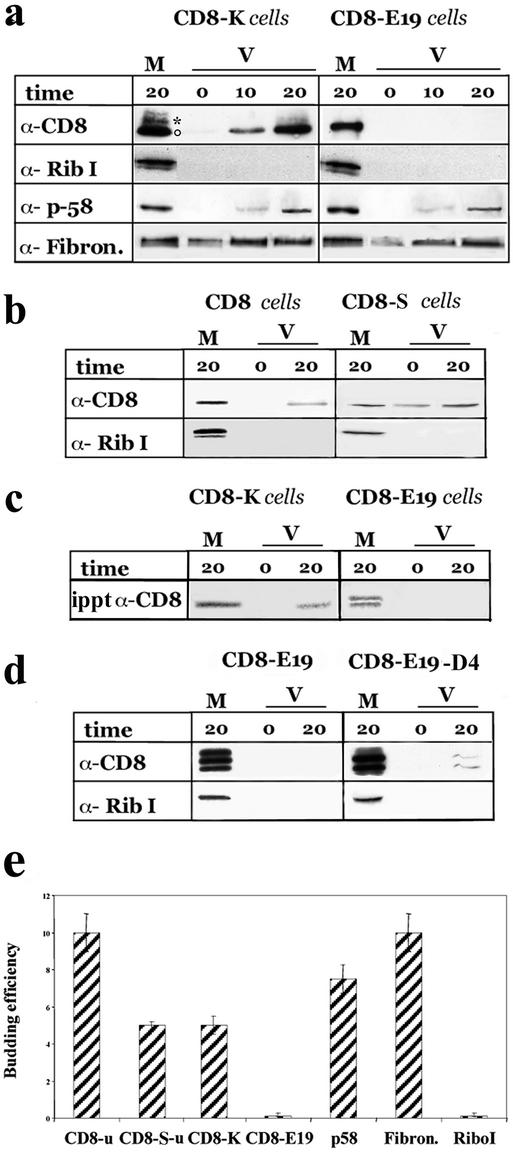
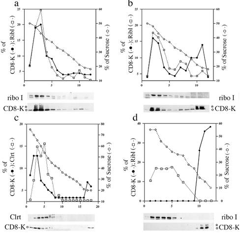
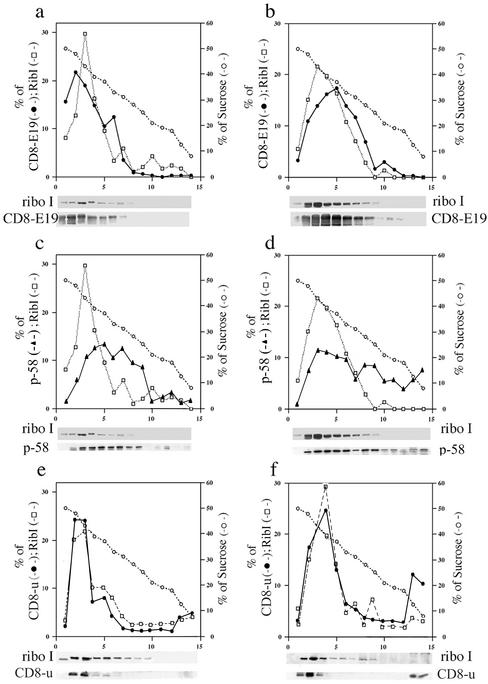
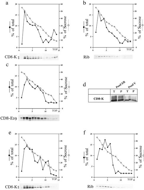
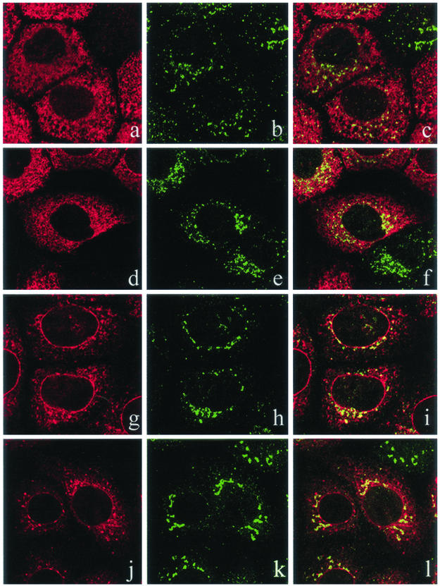
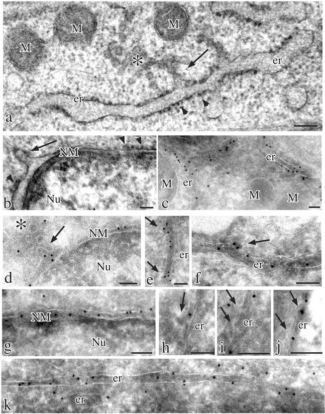
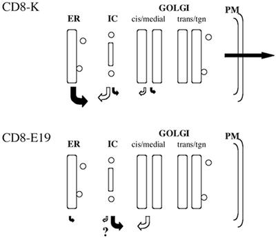
Many endoplasmic reticulum (ER) proteins maintain their residence by dynamic retrieval from downstream compartments of the secretory pathway. In previous work we compared the retrieval process mediated by the two signals, KKMP and KDEL, by appending them to the same neutral reporter protein, CD8, and found that the two signals determine a different steady-state localization of the reporter. CD8-K (the KDEL-bearing form) was restricted mainly to the ER, whereas CD8-E19 (the KKMP-bearing form) was distributed also to the intermediate compartment and Golgi complex. To investigate whether this different steady-state distribution reflects a difference in exit rates from the ER and/or in retrieval, we have now followed the first steps of export of the two constructs from the ER and their trafficking between ER and Golgi complex. Contrary to expectation, we find that CD8-K is efficiently recruited into transport vesicles, whereas CD8-E19 is not. Thus, the more restricted ER localization of CD8-K must be explained by a more efficient retrieval to the ER. Moreover, because most of ER resident CD8-K is not O-glycosylated but almost all CD8-E19 is, the results suggest that CD8-K is retrieved from the intermediate compartment, before reaching the Golgi, where O-glycosylation begins. These results illustrate how different retrieval signals determine different trafficking patterns and pose novel questions on the underlying molecular mechanisms.


Figures

Figure 1
Figure 1
CD8-K, but not CD8-E19, is efficiently exported from the ER in vitro. (a) Total microsomal fractions from cells stably expressing CD8-K or CDE19 were used to program budding assays in vitro as detailed in MATERIALS AND METHODS. The incubations were performed for the indicated times (in minutes; 0 indicates samples incubated on ice for 20 min) and then the microsomal (M) and the vesicular (V) fractions were separated by differential centrifugation and analyzed by SDS-PAGE and Western immunoblot to detect theindicated proteins. One-tenth of the M and 100% of the V fractions were loaded on the gels. The experiment shown is representative of seven and three independent assays for CD8-K and CD8-E19, respectively. ○ and * indicate the position on the gels of the nonglycosylated and initially glycosylated forms of CD8-K, respectively. (b) Total microsomal fraction from cells stably expressing CD8 and CD8-S were used for budding assay as described above. One-tenth of M and one entire V fraction, and one-half of M and three V fractions were loaded on the gels to detect CD8u and CD8-Su, respectively. Only the portions of the blots containing the nonglycosylated forms of the two proteins were analyzed. (c) Cells stably expressing CD8-K and CD8-E19 were radiolabeled for 20 min with [35S]cysteine and methionine and then used for the budding assay. The M and V fractions were lysed with 1% SDS, subjected to immunoprecipitation, and the total immunoprecipitated products were loaded on the gel. The dried gel was exposed for 3 d to a PhosphorImager (Bio-Rad, Hercules, CA) screen. (d) FRT cells were transiently transfected with pT8E19 or pT8D4, as described in MATERIALS AND METHODS and then used for the budding assay. One hundred percent of the M and V fractions were loaded on the gels. (e) Quantitation of budding efficiency of each protein recovered in the V fraction with respect to the total (M + V fractions). Only the results obtained with assays from stably expressing cells analyzed by immunoblotting are reported. Different exposures of the immunoblots were analyzed with the NIH Image program (see MATERIALS AND METHODS). The percentage of protein measured in the V fraction of incubations held on ice was subtracted from the corresponding incubations performed at 37°C. SD is indicated (n = 3).
Figure 2
Figure 2
Sucrose gradient analysis supports the evidence that CD8-K protein is exported from the ER to a vesicular fraction during the in vitro assay. See MATERIALS AND METHODS for details. Incubation mixtures of budding reactions programmed with microsomes from cells stably expressing CD8-K were examined by sedimentation on discontinuous sucrose gradients, except for c that reports the result of a flotation gradient. (a) Reaction mixture held on ice. (b–d) Reaction mixtures incubated for 20 min at 37°C. (d) At the end of the incubation, the microsomes were removed by centrifugation and the supernatant first mixed on ice with a corresponding amount of microsomes from parental FRT cells and then loaded on the top of the gradient. For all the gradients, equal aliquots from each fraction were analyzed by SDS-PAGE and Western immunoblot to detect the indicated proteins. The percentage of total protein contained in each fraction is reported on the left scale; on the right scale is the percentage (wt/vol) of sucrose. The relevant part of the immunoblot used for densitometry scanning is shown below each panel. ○ and * indicate the position on the gels of the nonglycosylated and initially glycosylated forms of CD8-K, respectively.
Figure 3
Figure 3
Sucrose gradient analysis confirms that CD8-E19 is not exported from the ER during the in vitro assay. Incubation mixtures of budding reactions programmed with microsomes from CD8-E19 (a–d), or from wild-type CD8 (e and f)-expressing cells, were examined by sedimentation on discontinuous sucrose gradients. (a, c, and e) Reaction mixtures held on ice. (b, d, and f) Reaction mixtures incubated for 20 min at 37°C. Couples a and c and b and d each refer to a single gradient. In e and f, only the portion of the blots containing the unglycosylated form of CD8 was analyzed. Gradient analysis and illustration as in Figure 2.
Figure 4
Figure 4
Cell fractionation analysis of postnuclear supernatant fractions obtained from cultured cells reveals that CD8-K has an higher rate of trafficking in vivo than CD8-E19. Postnuclear supernatant fractions were prepared from exponentially growing, subconfluent cultures of CD8-K– and CD8-E19–expressing cells and analyzed by sedimentation on discontinuous sucrose gradients. Couples a and b and e and f each refer to a single gradient and Western blot. (d) Fractions 2 and 3 (pool ER) and 13 and 14 (pool V) of a gradient like the one in a were pooled and divided in two equal aliquots; one aliquot (T), was incubated in 20% trichloroacetic acid and the precipitated proteins collected by centrifugation; the other (P) was diluted fourfold and centrifuged for 30 min at 45,000 rpm in a Beckman Coulter TL-100 centrifuge to recover the membrane fraction. Finally, the four pellets were analyzed by SDS-PAGE and Western immunoblot. (e and f) CD8-K–expressing cultures were incubated for 3 h at 15°C before being processed and analyzed as described above. Gradient analysis and illustration as in Figure 2.
Figure 5
Figure 5
A fraction of CD8-K, but not CD8-E19, moves to the IC when the cells are incubated at 15°C. Single optical sections of a confocal indirect immunofluorescence analysis. (a–c and g–i) CD8-K– and CD8-E19–expressing cells, respectively, incubated at 37°C. (d–f and j–l) CD8-K– and CD8-E19–expressing cells, respectively, incubated for 3 h at 15°C. Left, CD8-K or CD8-E19; middle, ERGIC-p58; and right, merge.
Figure 6
Figure 6
Different presence of CD8-K and CD8-E19 at ER exit sites in transiently transfected HuH-7 cells. Conventional thin section electron microscopy (a and b) and cryoimmunoelectron microscopy (c–k) of parental (a and b) and transiently transfected HuH-7 cells expressing CD8-K (c–f) and CD8-E19 (g–k) proteins. The ultrastructural analysis revealed the presence of long ER cisternae and outer nuclear membranes mostly studded with ribosomes (arrowheads in a and b) and occasionally showing smooth areas with emerging protrusions (arrows in a and b) facing IC units (asterisk in a). Immunogold labeling with anti-CD8 polyclonal antibody and protein A-colloidal gold conjugates were dense and unevenly distributed over ER cisternae (c), whereas the gold particles frequently were localized on the smooth ribosome-free areas of the membranes and clustered over protrusions extending from them (e and f, arrows). Similar clustering of gold particles was observed in protrusions extending from the outer nuclear membranes (d, arrow). Immunolabeling was also present on IC units (d, asterisk). In contrast, immunogold labeling of CD8-E19–expressing cells was homogeneously distributed over either outer nuclear membranes (g) or ER cisternae (k) and the protrusions extending from the ER (arrows in h–j) were often unlabeled but sometimes labeled (j). M, mitochondria; er, endoplasmic reticulum; NM, nuclear membrane; Nu, nucleus. Bars, 0.1 μm.
Figure 7
Figure 7
Model of the trafficking events of CD8-K and CD8-E19. The size of the arrows indicates the relative rate of the transport event considered. Question mark indicates the possibility that CD8-E19 is retrieved at low rate also from the IC, an event yet to be proven.

Similar articles

Cited by

References

    1. Andersson H, Kappeler F, Hauri HP. Protein targeting to endoplasmic reticulum by dilysine signals involves direct retention in addition to retrieval. J Biol Chem. 1999;274:15080–15084. - PubMed
    1. Aoe T, Lee AJ, van Donselaar E, Peters PJ, Hsu VW. Modulation of intracellular transport by transported proteins: insight from regulation of COPI-mediated transport. Proc Natl Acad Sci USA. 1998;95:1624–1629. - PMC - PubMed
    1. Bannykh SI, Rowe T, Balch WE. The organization of endoplasmic reticulum export complexes. J Cell Biol. 1996;135:19–35. - PMC - PubMed
    1. Bonatti S, Migliaccio G, Simons K. Palmitylation of viral membrane glycoproteins takes place after exit from the endoplasmic reticulum. J Biol Chem. 1989;264:12590–12595. - PubMed
    1. Cosson P, Demolliere C, Hennecke S, Duden R, Letourneur F. δ- and ξ-COP, two coatomer subunits homologous to clathrin-associated proteins, are involved in ER retrieval. Embo J. 1996;15:1792–1798. - PMC - PubMed

Publication types