. 2003 Mar;14(3):926-38.
doi: 10.1091/mbc.e02-09-0573.

HIV-1 envelope proteins complete their folding into six-helix bundles immediately after fusion pore formation

Affiliations

HIV-1 envelope proteins complete their folding into six-helix bundles immediately after fusion pore formation

Ruben M Markosyan et al. Mol Biol Cell. 2003 Mar.

Abstract

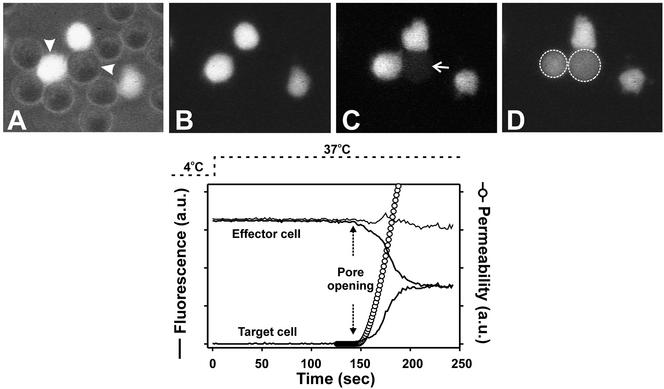
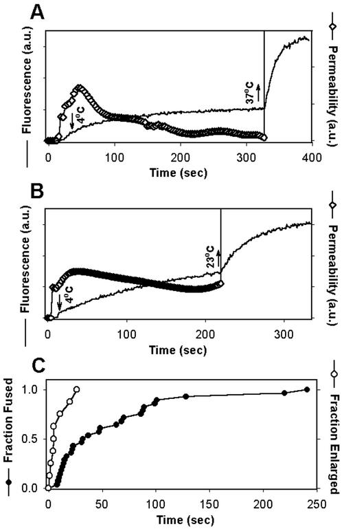
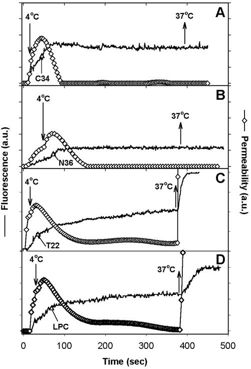
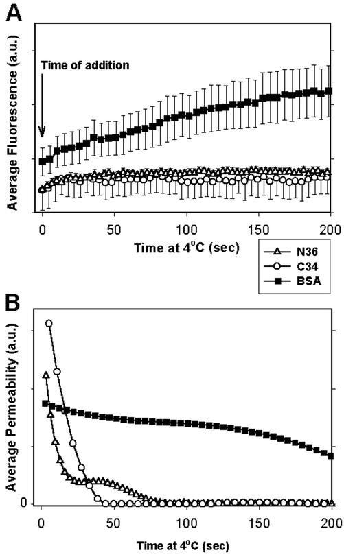
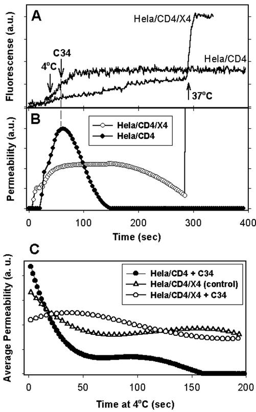
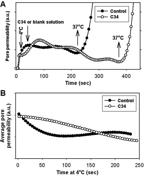
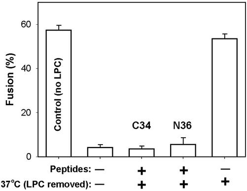
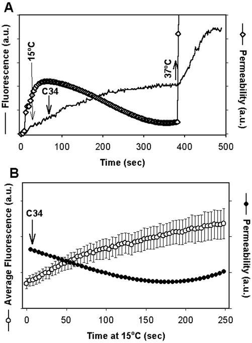
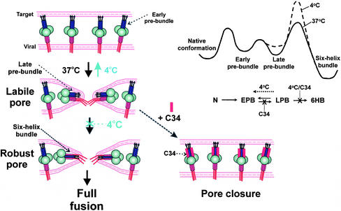
Fusion proteins of many viruses, including HIV-1 envelope protein (Env), fold into six-helix bundle structures. Fusion between individual Env-expressing cells and target cells was studied by fluorescence microscopy, and a temperature jump technique, to determine whether folding of Env into a bundle is complete by the time fusion pores have formed. Lowering temperature to 4 degrees C immediately after a pore opened halted pore growth, which quickly resumed when temperature was raised again. HIV gp41-derived peptides that inhibit bundle formation (C34 or N36) caused the cold-arrested pore to quickly and irreversibly close, demonstrating that bundle formation is not complete by the time a pore has formed. In contrast, lowering the temperature to an intermediate value also halted pore growth, but the pore was not closed by the bundle-inhibiting peptides, and it enlarged when temperature was again elevated. This latter result shows that bundle formation is definitely required for the fusion process, but surprisingly, some (if not all) bundle formation occurs after a pore has formed. It is concluded that an essential function of the bundle is to stabilize the pore against collapse and ensure its growth.


Figures

Figure 1
Figure 1
Monitoring fusion pore dynamics through fluorescent dye redistribution between effector and target cells. Top panel: (A) Effector (TF228.1.16) cells labeled with calcein and unlabeled target (HeLaT4+) cells were coincubated for 3 h at 23°C. These cells were then placed in an experimental chamber maintained at 4°C. The image of A is a superposition of fluorescence and phase contrast after placement. Fusion was then triggered by quickly raising the temperature to 37°C and monitored by the transfer of calcein from the effector cell to the target cell (marked by arrowheads). (B–D) The onset of dye movement is shown in C (arrow) and after full redistribution in D (the dotted circles mark the region of interest encompassing the effector and the target cell). Bottom panel: Changes in mean fluorescence intensity of the target (lower thick solid line) and the effector (upper thick solid line) cell with time after raising temperature to 37°C. The temperature profile is shown by dotted line. The thin solid line is the sum of the fluorescence intensities of the effector and target cells. Once the fusion pore formed (marked by vertical dotted arrows), it took ∼1 min for the calcein to fully redistribute. The pore permeability profile in arbitrary units (open circles) was calculated from fluorescence intensity changes using Eq. (1) (see MATERIALS AND METHODS).
Figure 2
Figure 2
Arresting the fusion pore by low temperature. Immediately after a fusion pore formed between TF228.1.16 and HeLaT4+ cells, pore growth was arrested by quickly reducing the temperature from 37 to 4°C (downward arrows in A and B). The fluorescence intensities of the target cell over time are shown by solid lines. (A and B) Upward arrows denote raising temperature back to 37°C (A) or to 23°C (B). The pore permeability (⋄) was calculated from the fluorescence traces using Eq. (2). After elevating the temperature from 4°C, the pore rapidly enlarged (nearly vertical lines in A and B). (C) The waiting time distribution between the fast increase of temperature at TAS and formation of a fusion pore, plotted as the fraction of cells that fused (●) was significantly longer than the distribution for time between exposing the cold-arrested pores to 37°C and their rapid enlargement, plotted as the fraction of pores that enlarged (○).
Figure 3
Figure 3
Cold-arrested fusion pores were closed by peptides that block 6HB formation but not by other inhibitors of fusion. The time at which the temperature was dropped to 4°C to block pore growth is marked by downward arrows in each panel. The fluorescence intensities of target cells (HeLaT4+, solid lines) were used to calculate pore permeability from Eq. (2) (⋄). Once pore growth was blocked at 4°C, inhibitory agents were added (indicated by ▵), allowed to bind for ∼5 min, and the temperature was then raised back to 37°C (upward arrows). The inhibitors were (A) 47 nM C34 peptide, (B) 4 μM N36 peptide, (C) 0.4 μg/ml T22 peptide, and (D) 0.2 mg/ml LPC.
Figure 4
Figure 4
Average rate of pore closure after adding inhibitors of 6HB formation. The fluorescence (A) and pore permeability (B) after the addition (indicated by downward arrow) at 4°C of the inhibitory peptides C34 (47 nM, ○, n = 5) or N36 (4 μM, ▵, n = 7) or, as a control, BSA (▪, n = 8), as averaged from multiple experiments. In the control experiments, we changed the external solution by the same procedure used to add peptides, to ensure that the peptides themselves were causing the inhibitory effect, rather than a simple shear stress on the E/T cell pairs. (A) Fluorescence traces from individual experiments were aligned at the time of peptide addition, and the average fluorescence from multiple experiments over the next 200 s was calculated. Bars, SEs of mean. (B) Pore permeabilities, calculated from the averaged fluorescence intensities (traces in A). The addition of C34 or N36, but not BSA, caused pore closure.
Figure 5
Figure 5
Cold-arrested pores for target cells expressing a high level of CXCR4 were robust, whereas those expressing a low level of CXCR4 were labile. HeLa cells were transfected with either CD4- and CXCR4-bearing plasmids (HeLa/CD4/X4, high CXCR4) or a CD4-bearing plasmid alone (HeLa/CD4, low CXCR4). Immediately after arresting pore growth by lowering temperature to 4°C, 150 nM of C34 peptide was added to the extracellular solution. (A) Fluorescence intensity changes of transfected HeLa cells. (B) Pore permeability profiles calculated from the traces in A. (C) Average permeability as a function of time at 4°C for HeLa/CD4/X4 cells with (○, n = 6) and without (▵, n = 5) C34 peptide, and for HeLa/CD4 cells in the presence of C34 peptide (●, n = 5). Average pore permeability was calculated as described in the legend to Figure 4.
Figure 6
Figure 6
The cold-arrested pore is robust for 3T3. T4. CXCR4 cells as targets. (A) C34 (150 nM, ○) or a blank solution (●) was added at 4°C to cold-arrested fusion pores formed between TF228.1.16 and 3T3.T4.CXCR4 cells. (B) Average pore permeability profiles at 4°C in the presence (○) and in absence (●) of C34 peptide.
Figure 7
Figure 7
Peptides that prevent the formation of six-helix bundles still inhibit fusion when added at an LPC-arrested stage for high CXCR4-expressing cells. TF228.1.16 cells were coincubated with 3T3.T4.CXCR4 cells for 3 h at 23°C followed by incubation at 37°C. The full extent of fusion is indicated (first bar). A lipid-arrested stage (LAS) of fusion was created by adding 0.3 mg/ml LPC to cells for 3 min at 23°C, raising temperature to 37°C for 15 min and then removing LPC by repeated washings with a BSA-containing solution at 23°C. The extent of fusion was negligible (second bar). The addition of 150 nM C34 (third bar) or 4 μM N36 (fourth bar) at LAS abolished any additional fusion when temperature was raised to 37°C, showing that 6HBs have not yet formed. When peptides were not added after creating LAS, raising temperature to 37°C led to full extent of fusion (fifth bar). Bars, SEs of mean (n = 4).
Figure 8
Figure 8
Pores arrested at 15°C are robust for target cells expressing low levels of CXCR4. (A) Fluorescence trace of calcein accumulation within the target cells (solid line). Temperature was lowered from 37 to 15°C (long downward arrow) and 150 nM C34 peptide was then added, as indicated. The pore permeability (⋄) was calculated from the curve of fluorescence intensity using Eq. (2). The pore fully enlarged (nearly a vertical line) immediately after temperature was raised back to 37°C (upward arrow). (B) The average pore permeability (●) calculated from the average fluorescence profile (○) as a function of time at 4°C in the presence of C34. HeLaT4+ cells were used as targets. Bars, SEs of mean (n = 5).
Figure 9
Figure 9
A proposed mechanism for the conformational changes of gp41 in pore formation and stabilization. Left panels: Conversion of Env in early prebundles (top) at a fusion site into late prebundles (middle) creates a labile pore, and subsequent folding into the final six-helix bundle configuration establishes the robust pore (bottom). The gp120 is depicted as large circles; N-terminal helices (purple rectangles) of gp41 are adjacent to the fusion peptides (arrows) and C-terminal helices (red rectangles) are adjacent to the membrane-spanning domain (this domain and cytoplasmic regions are solid red lines). Early prebundles are shown as an extended conformation with N- and C-terminal helices well separated from each other; late prebundles are depicted with one C-terminal helix bound into a groove of the triple-stranded coiled-coil. Arresting a labile pore by 4°C leads to reduction in pore size and eventual closure (transition from middle to top). The addition of 6HB blockers (C34, red rectangle) to the labile pore results in rapid pore closure (dashed arrow). Late prebundles stabilize the pore, but only when the six-helix bundle is formed is the pore permanently stabilized against closure. Top right: The proposed energy profile of gp41 during the fusion reaction at 37°C (solid line) and at 4°C (dashed line). In the reaction scheme, N stands for the native conformation, EPB and LPB are early and late prebundle conformations, respectively. Because the formation of the 6HB should be constrained by limitations in protein reorientation, its activation barrier would be controlled by entropy whose energetic consequences are more sensitive to temperature than is enthalpy. The transition from late prebundles to bundles is therefore expected to be more temperature-sensitive than the other transitions.

Similar articles

Cited by

References

    1. Cao J, Bergeron L, Helseth E, Thali M, Repke H, Sodroski J. Effects of amino acid changes in the extracellular domain of the human immunodeficiency virus type 1 gp41 envelope glycoprotein. J Virol. 1993;67:2747–2755. - PMC - PubMed
    1. Chan DC, Chutkowski CT, Kim PS. Evidence that a prominent cavity in the coiled coil of HIV type 1 gp41 is an attractive drug target. Proc Natl Acad Sci USA. 1998;95:15613–15617. - PMC - PubMed
    1. Chan DC, Kim PS. HIV entry and its inhibition. Cell. 1998;93:681–684. - PubMed
    1. Chen CH, Matthews TJ, McDanal CB, Bolognesi DP, Greenberg ML. A molecular clasp in the human immunodeficiency virus (HIV) type 1 TM protein determines the anti-HIV activity of gp41 derivatives: implication for viral fusion. J Virol. 1995;69:3771–3777. - PMC - PubMed
    1. Chernomordik L, Kozlov MM, Zimmerberg J. Lipids in biological membrane fusion. J Membr Biol. 1995;146:1–14. - PubMed

Publication types

LinkOut - more resources