. 2003 Mar;14(3):939-57.
doi: 10.1091/mbc.e02-10-0633.

Peroxisome biogenesis occurs in an unsynchronized manner in close association with the endoplasmic reticulum in temperature-sensitive Yarrowia lipolytica Pex3p mutants

Affiliations

Peroxisome biogenesis occurs in an unsynchronized manner in close association with the endoplasmic reticulum in temperature-sensitive Yarrowia lipolytica Pex3p mutants

Roger A Bascom et al. Mol Biol Cell. 2003 Mar.

Abstract

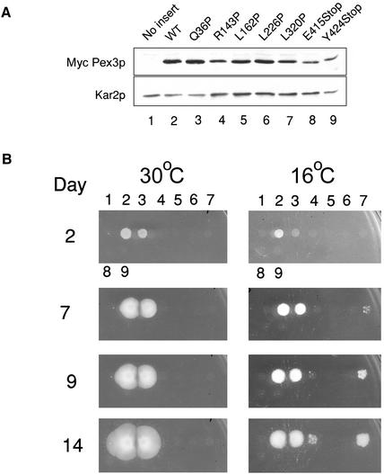
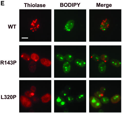
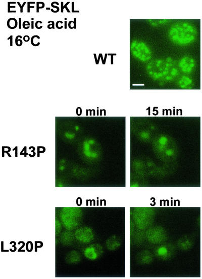
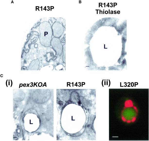
Pex3p is a peroxisomal integral membrane protein required early in peroxisome biogenesis, and Pex3p-deficient cells lack identifiable peroxisomes. Two temperature-sensitive pex3 mutant strains of the yeast Yarrowia lipolytica were made to investigate the role of Pex3p in the early stages of peroxisome biogenesis. In glucose medium at 16 degrees C, these mutants underwent de novo peroxisome biogenesis and exhibited early matrix protein sequestration into peroxisome-like structures found at the endoplasmic reticulum-rich periphery of cells or sometimes associated with nuclei. The de novo peroxisome biogenesis seemed unsynchronized, with peroxisomes occurring at different stages of development both within cells and between cells. Cells with peripheral nascent peroxisomes and cells with structures morphologically distinct from peroxisomes, such as semi/circular tubular structures that immunostained with antibodies to peroxisomal matrix proteins and to the endoplasmic reticulum-resident protein Kar2p, and that surrounded lipid droplets, were observed during up-regulation of peroxisome biogenesis in cells incubated in oleic acid medium at 16 degrees C. These structures were not detected in wild-type or Pex3p-deficient cells. Their role in peroxisome biogenesis remains unclear. Targeting of peroxisomal matrix proteins to these structures suggests that Pex3p directly or indirectly sequesters components of the peroxisome biogenesis machinery. Such a role is consistent with Pex3p overexpression producing cells with fewer, larger, and clustered peroxisomes.


Figures

Figure 1
Figure 1
PTS1- and PTS2-containing matrix proteins are mistargeted to the cytosol in Y. lipolytica cells mutant for Pex3p. (A) Wild-type (WT) strain E122, original mutant strain mut12-1, and transformed strain M12TR were grown at 30°C in glucose-containing YND medium, transferred to oleic acid-containing YNO medium, and incubated at 30°C for an additional 8 h. Cells were processed for immunofluorescence microscopy with rabbit antibodies to the PTS1 tripeptide SKL and guinea pig antibodies to the PTS2-targeted enzyme thiolase (THI). Rabbit primary antibodies were detected with fluorescein-conjugated goat anti-rabbit immunoglobulin G secondary antibodies, and guinea pig primary antibodies were detected with rhodamine-conjugated donkey anti-guinea pig immunoglobulin G secondary antibodies. Bar, 5 μm. (B) Nucleotide sequence of the YlPEX3 gene and deduced amino acid sequence of YlPex3p. Translation was shown experimentally to occur from the indicated ATG rather than from an upstream, inframe ATG. These sequence data have been deposited in the DDBJ/EMBL/GenBank databases under accession number AF474003. (C) Sequence similarity between YlPex3p and Pex3 proteins of other organisms. Numeric scale at top indicates protein length in amino acids. Colorimetric scale of protein similarity is defined at right. The five blocks of amino acid sequence conserved among the different Pex3 proteins as defined by the program Macaw2, and the amino acid changes made to generate ts mutant forms of Pex3p, are presented at bottom. (D) Ultrastructure of wild-type and pex3KOA cells. The wild-type (WT) strain E122 and the pex3KOA mutant strain were grown in YPD medium and then incubated for 8 h at 30°C in YPBO medium. Cells were fixed in 1.5% KMnO4 and processed for electron microscopy. Peroxisomes (P) were observed in wild-type cells but not in pex3KOA cells, which instead contained numerous small vesicles (V). L, lipid droplet; M, mitochondrion, N, nucleus. Bar, 0.5 μm.
Figure 2
Figure 2
Pex3p is a peroxisomal integral membrane protein exposed to the cytosol. (A) Pex3p is localized to peroxisomes. Wild-type E122 cells incubated in oleic acid-containing YPBO medium for 8 h were processed for immunofluorescence microscopy with guinea pig primary antibodies to Pex3p and rabbit primary antibodies to the PTS1 tripeptide SKL. Primary antibodies were detected as described in the legend to Figure 1A. Bar, 2.5 μm. (B) Pex3p exhibits properties of an integral membrane protein. A functionally complementing form of Pex3p tagged with the c-Myc epitope (MycPex3p) resists extraction from a 20KgP subcellular fraction by treatment with 1.0 M NaCl, 1.0 M urea, and 0.1 M Na2CO3, pH 11. (C) Pex3p is exposed to the cytosol. MycPex3p in a 20KgP subcellular fraction is sensitive to exogenously added trypsin in both the absence and the presence of the detergent Triton X-100. In contrast, the peroxisomal matrix protein thiolase (THI) is resistant to exogenously added trypsin in the absence of detergent and sensitive in the presence of detergent.
Figure 3
Figure 3
Temperature-sensitive growth of Pex3p variants. (A) Levels of the wild-type and mutant c-Myc-tagged Pex3 proteins expressed in transformed pex3KOA cells are comparable. “No insert” refers to the pex3KOA strain containing the vector pINA445 alone. Lysates of the various strains were subjected to immunoblotting with the 9E10 antibody against the c-Myc epitope. The immunodetection of Kar2p was used as a loading control. (B) Mutant MycPex3 proteins R143P and L320P confer ts growth on oleic acid-containing agar to the pex3KOA strain. The days of incubation at 30°C or 16°C are indicated. Numbering of strains is as in A.
Figure 4
Figure 4
Peroxisome biogenesis occurs in cells grown in glucose-containing medium at 16°C. (A) Cells of strains able to grow on oleic acid-containing YNO agar plates at 30°C (wild-type [WT] and MycPex3p-Q36P) localized thiolase to peroxisomes by immunofluorescence, whereas cells of strains unable to grow on YNO agar plates at 30°C (MycPex3p-R143P, -L162P, -L226P, -L320P, -E415Stop, and -Y424Stop) showed a predominantly cytosolic localization for thiolase, upon incubation in oleic acid-containing YNO medium at 30°C. (B) At 16°C in glucose-containing YND medium, thiolase was targeted to peroxisomes of the WT strain and of the strain expressing MycPex3p-Q36P; however, targeting was less efficient in the latter strain. Strains expressing MycPex3p-L162P, -L226P, -E415Stop, and -Y424Stop showed a cytosolic localization for thiolase, whereas thiolase sequestration was first detected in MycPex3p-R143P and MycPex3p-L320P at one site, or sometimes at multiple sites, in the peripheral regions of cells.
Figure 5
Figure 5
De novo peroxisome biogenesis occurs in cells grown in glucose-containing medium in regions associated with ER elements. (A) Immunofluorescence analysis demonstrating that the peroxisome-like structures found at the periphery of glucose-grown MycPex3p-R143p– or MycPex3p-L320P–expressing cells contain the matrix enzyme AOX but not the matrix enzyme thiolase, suggesting that these structures are nascent peroxisomes undergoing biogenesis. Bar, 3 μm. (B) Peripherally located peroxisomes contain different matrix proteins. Y. lipolytica cells were analyzed by immunofluorescence with antibodies to thiolase, the PTS1 SKL, AOX, and ICL. Bar, 3 μm. (C) Nascent, peripherally located peroxisomes are found in ER-rich regions of the cell and sometimes associated with the nuclear envelope. Cells were processed for immunofluorescence and doubly labeled with antibodies to thiolase and to the ER resident protein Kar2p. Bar, 3 μm. (D) Individual, peripherally located peroxisomes are often positioned close to nuclei. Cells were labeled with antibodies to thiolase and with the nuclear stain, DAPI. Bar, 3 μm. (E) Peroxisomes are randomly positioned with respect to lipid droplets in glucose-grown cells. Cells were labeled with antibodies to thiolase and with the neutral lipid stain BODIPY 493/503. Bar, 3 μm. (F) Peroxisome-like structures (P) are observed by electron microscopy closely associated with ER elements (E) in cells undergoing de novo peroxisome biogenesis. Cells were grown in glucose-containing YND medium at 16°C for 48 h.
Figure 5
Figure 5
De novo peroxisome biogenesis occurs in cells grown in glucose-containing medium in regions associated with ER elements. (A) Immunofluorescence analysis demonstrating that the peroxisome-like structures found at the periphery of glucose-grown MycPex3p-R143p– or MycPex3p-L320P–expressing cells contain the matrix enzyme AOX but not the matrix enzyme thiolase, suggesting that these structures are nascent peroxisomes undergoing biogenesis. Bar, 3 μm. (B) Peripherally located peroxisomes contain different matrix proteins. Y. lipolytica cells were analyzed by immunofluorescence with antibodies to thiolase, the PTS1 SKL, AOX, and ICL. Bar, 3 μm. (C) Nascent, peripherally located peroxisomes are found in ER-rich regions of the cell and sometimes associated with the nuclear envelope. Cells were processed for immunofluorescence and doubly labeled with antibodies to thiolase and to the ER resident protein Kar2p. Bar, 3 μm. (D) Individual, peripherally located peroxisomes are often positioned close to nuclei. Cells were labeled with antibodies to thiolase and with the nuclear stain, DAPI. Bar, 3 μm. (E) Peroxisomes are randomly positioned with respect to lipid droplets in glucose-grown cells. Cells were labeled with antibodies to thiolase and with the neutral lipid stain BODIPY 493/503. Bar, 3 μm. (F) Peroxisome-like structures (P) are observed by electron microscopy closely associated with ER elements (E) in cells undergoing de novo peroxisome biogenesis. Cells were grown in glucose-containing YND medium at 16°C for 48 h.
Figure 6
Figure 6
Peroxisomal matrix proteins are present in semi/circular tubular structures in Pex3p ts mutants undergoing peroxisome proliferation in oleic acid-containing medium. Cells were grown in glucose-containing YND medium for 48 h at 16°C and then incubated in oleic acid-containing YNO medium for 8 h at 16°C. (A) Immunofluorescence analysis with antibodies to thiolase showing that MycPex3p-R143P– and MycPex3p-L320P–expressing cells incubated in oleic acid-containing (YNO) medium at 16°C contain both peripherally located peroxisomes and distinct semi/circular tubular structures that immunostain for thiolase. (B) The semi/circular tubular structures immunopositive for thiolase do not surround nuclei. Nuclei were detected by staining with DAPI. (C) Semi/circular tubular structures immunopositive for thiolase surround lipid droplets. Lipid droplets were detected by staining with BODIPY 493/503. (D) Different peroxisomal matrix proteins are present in the semi/circular tubular structures. Immunofluorescence analysis showing colocalization of signals for antibodies to the PTS1 SKL (i), AOX (ii), and ICL (iii) with the signal for antibodies to thiolase in semi/circular tubular structures. (E) Immunofluorescence analysis showing the ER resident protein Kar2p partially colocalizes with thiolase in semi/circular tubular structures. (F) Immunofluorescence analysis shows no colocalization of thiolase and the vacuole resident protein carboxypeptidase Y (CPY), indicating that the semi/circular tubular structures are not vacuoles in the process of degrading peroxisomes. Bars, 3 μm.
Figure 6
Figure 6
Peroxisomal matrix proteins are present in semi/circular tubular structures in Pex3p ts mutants undergoing peroxisome proliferation in oleic acid-containing medium. Cells were grown in glucose-containing YND medium for 48 h at 16°C and then incubated in oleic acid-containing YNO medium for 8 h at 16°C. (A) Immunofluorescence analysis with antibodies to thiolase showing that MycPex3p-R143P– and MycPex3p-L320P–expressing cells incubated in oleic acid-containing (YNO) medium at 16°C contain both peripherally located peroxisomes and distinct semi/circular tubular structures that immunostain for thiolase. (B) The semi/circular tubular structures immunopositive for thiolase do not surround nuclei. Nuclei were detected by staining with DAPI. (C) Semi/circular tubular structures immunopositive for thiolase surround lipid droplets. Lipid droplets were detected by staining with BODIPY 493/503. (D) Different peroxisomal matrix proteins are present in the semi/circular tubular structures. Immunofluorescence analysis showing colocalization of signals for antibodies to the PTS1 SKL (i), AOX (ii), and ICL (iii) with the signal for antibodies to thiolase in semi/circular tubular structures. (E) Immunofluorescence analysis showing the ER resident protein Kar2p partially colocalizes with thiolase in semi/circular tubular structures. (F) Immunofluorescence analysis shows no colocalization of thiolase and the vacuole resident protein carboxypeptidase Y (CPY), indicating that the semi/circular tubular structures are not vacuoles in the process of degrading peroxisomes. Bars, 3 μm.
Figure 7
Figure 7
Semi/circular tubular structures give rise to peroxisome-like structures in living cells. Cells expressing the chimeric protein EYFP-SKL and either MycPex3p-R143P or MycPex3p-L320P were grown at 16°C for 48 h in glucose-containing YND medium and then incubated for 8 h in oleic acid-containing YNO medium. EYFP-SKL is imported into peroxisomes in wild-type (WT) cells. In cells coexpressing MycPex3p-R143P or MycPex3p-L320P, the EYFP-SKL protein is targeted to the semi/circular tubular compartment at 0 min, which give rises to peroxisome-like structures by 15 min. Bar, 3 μm.
Figure 8
Figure 8
Microscopic analysis of peroxisomes in cells expressing MycPex3p-R143P and of structures surrounding lipid droplets. Cells were grown for 48 h in glucose-containing YND medium at 16°C, transferred to oleic acid-containing YNO medium, and incubated for a further 8 h at 16°C. (A) Electron micrograph of peroxisomes (P) in MycPex3p-R143P–expressing cells. (B) Electron micrograph of the structure surrounding a lipid droplet (L) in cells expressing MycPex3p-R143P. The structure shows immunogold labeling with antibody to thiolase. (C, i) Electron micrographs of structures surrounding lipid droplets (L) in the pex3KOA strain (left) and the pex3KOA strain expressing MycPex3p-R143P (right). The structures are distinct from the lipid droplets themselves, with their phospholipid bilayer membranes seen to be wrapping around the phospholipid monolayer of the lipid droplets. The structure around the lipid droplet of the strain expressing MycPex3p-R143P contains a bulge (arrow), which is peroxisome-like in appearance. (ii) A semi/circular tubular structure immunostained for thiolase and surrounding a lipid droplet detected with BODIPY 493/503 resembles the structure surrounding a lipid droplet observed by electron microscopy of a MycPex3p-R143P-expressing cell seen in Figure 8B. Bar, 1.0 μm. Bars in electron micrographs, 0.1 μm.
Figure 9
Figure 9
Overexpression of Pex3p produces a reduced number of enlarged and clustered peroxisomes. (A) Immunoblot with 9E10 antibody of MycPex3p expressed from the native PEX3 promoter in the plasmid pINA445 (pINA445Pex3p) and overexpressed from the thiolase promoter (TC3Pex3p) in the wild-type E122 strain. Immunodetection of glucose-6-phosphate dehydrogenase (G6PDH) was done as a loading control. (B) Overexpressed MycPex3p is detected in the perinuclear/juxtanuclear region (i), as well as in semicircular structures on the surface of lipid droplets (ii). Wild-type cells overexpressing MycPex3p were analyzed by immunofluorescence with an affinity-purified antibody to Pex3p and with antibodies to the ER resident protein Kar2p (i). Nuclei were detected by staining with DAPI (i), and lipid droplets were detected by staining with BODIPY 493/503 (ii). Bars, 3 μm. (C) Wild-type E122 and original mut12-1 mutant cells overexpressing MycPex3p have reduced numbers of enlarged peroxisomes that exhibit clustering. Peroxisomes were detected by immunofluorescence with anti-thiolase antibodies. Nuclei were detected by staining with DAPI. Bar, 3 μm. (D) Electron micrographs of the wild-type (WT) E122 strain (left) and of the wild-type (middle) and the original mut12-1 (right) strains overexpressing MycPex3p. Overexpression of MycPex3p produces a reduced number of enlarged peroxisomes that cluster. L, lipid droplet; N, nucleus; P, peroxisome. Bar, 0.5 μm.
Figure 9
Figure 9
Overexpression of Pex3p produces a reduced number of enlarged and clustered peroxisomes. (A) Immunoblot with 9E10 antibody of MycPex3p expressed from the native PEX3 promoter in the plasmid pINA445 (pINA445Pex3p) and overexpressed from the thiolase promoter (TC3Pex3p) in the wild-type E122 strain. Immunodetection of glucose-6-phosphate dehydrogenase (G6PDH) was done as a loading control. (B) Overexpressed MycPex3p is detected in the perinuclear/juxtanuclear region (i), as well as in semicircular structures on the surface of lipid droplets (ii). Wild-type cells overexpressing MycPex3p were analyzed by immunofluorescence with an affinity-purified antibody to Pex3p and with antibodies to the ER resident protein Kar2p (i). Nuclei were detected by staining with DAPI (i), and lipid droplets were detected by staining with BODIPY 493/503 (ii). Bars, 3 μm. (C) Wild-type E122 and original mut12-1 mutant cells overexpressing MycPex3p have reduced numbers of enlarged peroxisomes that exhibit clustering. Peroxisomes were detected by immunofluorescence with anti-thiolase antibodies. Nuclei were detected by staining with DAPI. Bar, 3 μm. (D) Electron micrographs of the wild-type (WT) E122 strain (left) and of the wild-type (middle) and the original mut12-1 (right) strains overexpressing MycPex3p. Overexpression of MycPex3p produces a reduced number of enlarged peroxisomes that cluster. L, lipid droplet; N, nucleus; P, peroxisome. Bar, 0.5 μm.

References

    1. Aitchison JD, Szilard RK, Nuttley WM, Rachubinski RA. Antibodies directed against a yeast carboxyl-terminal peroxisomal targeting signal specifically recognize peroxisomal proteins from various yeasts. Yeast. 1992;8:721–734. - PubMed
    1. Ausubel FJ, Brent R, Kingston RE, Moore DD, Seidman JG, Smith JA, Struhl K. Current Protocols in Molecular Biology. New York: John Wiley & Sons; 1994.
    1. Blanchette-Mackie EJ, Dwyer NK, Barber T, Coxey RA, Takeda T, Rondinone CM, Theodorakis JL, Greenberg AS, Londos C. Perilipin is located on the surface layer of intracellular lipid droplets in adipocytes. J Lipid Res. 1995;36:1211–1226. - PubMed
    1. Brown DA. Lipid droplets: proteins floating on a pool of fat. Curr Biol. 2001;11:R446–R449. - PubMed
    1. Chang CC, South S, Warren D, Jones J, Moser AB, Moser HW, Gould SJ. Metabolic control of peroxisome abundance. J Cell Sci. 1999;112:1579–1590. - PubMed

Publication types

MeSH terms