. 2003 Mar;14(3):1027-42.
doi: 10.1091/mbc.e02-08-0503.

Loss of caveolin-1 gene expression accelerates the development of dysplastic mammary lesions in tumor-prone transgenic mice

Affiliations

Loss of caveolin-1 gene expression accelerates the development of dysplastic mammary lesions in tumor-prone transgenic mice

Terence M Williams et al. Mol Biol Cell. 2003 Mar.

Abstract

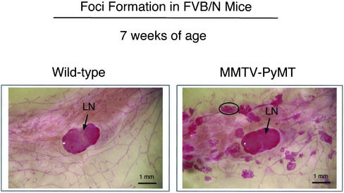
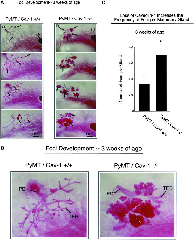
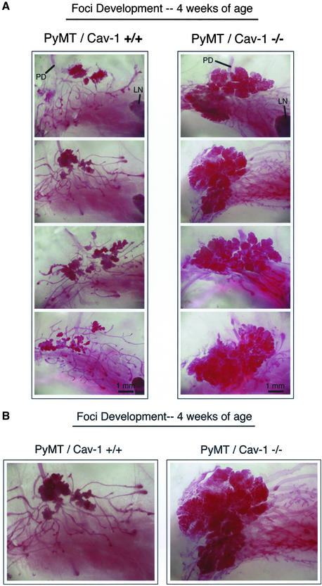
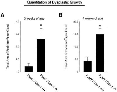
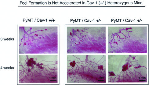
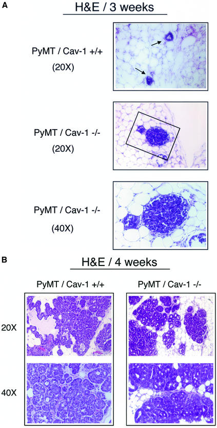
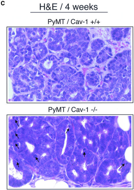
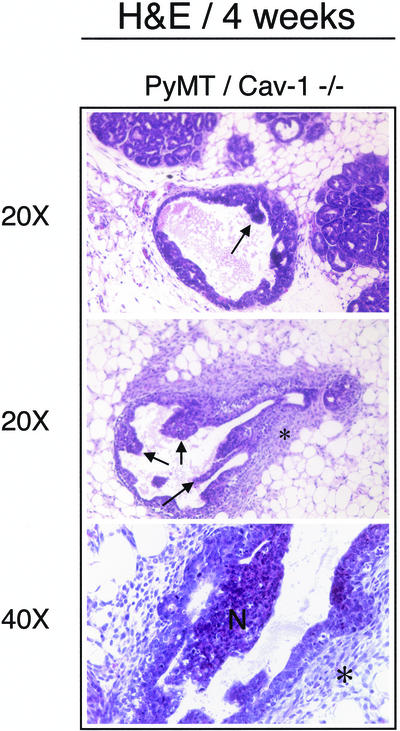
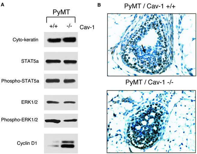
Caveolin-1 is the principal structural component of caveolae microdomains, which represent a subcompartment of the plasma membrane. Several independent lines of evidence support the notion that caveolin-1 functions as a suppressor of cell transformation. For example, the human CAV-1 gene maps to a suspected tumor suppressor locus (D7S522/7q31.1) that is frequently deleted in a number of carcinomas, including breast cancers. In addition, up to 16% of human breast cancers harbor a dominant-negative mutation, P132L, in the CAV-1 gene. Despite these genetic associations, the tumor suppressor role of caveolin-1 still remains controversial. To directly assess the in vivo transformation suppressor activity of the caveolin-1 gene, we interbred Cav-1 (-/-) null mice with tumor-prone transgenic mice (MMTV-PyMT) that normally develop multifocal dysplastic lesions throughout the entire mammary tree. Herein, we show that loss of caveolin-1 gene expression dramatically accelerates the development of these multifocal dysplastic mammary lesions. At 3 wk of age, loss of caveolin-1 resulted in an approximately twofold increase in the number of lesions (foci per gland; 3.3 +/- 1.0 vs. 7.0 +/- 1.2) and an approximately five- to sixfold increase in the total area occupied by these lesions. Similar results were obtained at 4 wk of age. However, complete loss of caveolin-1 was required to accelerate the appearance of these dysplastic mammary lesions, because Cav-1 (+/-) heterozygous mice did not show any increases in foci development. We also show that loss of caveolin-1 increases the extent and the histological grade of these mammary lesions and facilitates the development of papillary projections in the mammary ducts. Finally, we demonstrate that cyclin D1 expression levels are dramatically elevated in Cav-1 (-/-) null mammary lesions, consistent with the accelerated appearance and growth of these dysplastic foci. This is the first in vivo demonstration that caveolin-1 can function as a transformation suppressor gene.


Figures

Figure 1
Figure 1
Whole-mount analysis of the mammary glands of wild-type and PyMT mice in the FVB/N background. At 7 wk of age, the right fourth (inguinal) mammary glands were excised from virgin female mice, spread onto glass slides, fixed, and stained with carmine alum. Note the widespread appearance of multifocal dysplastic foci in PyMT FVB/N mice. A single dysplastic lesion is encircled. Arrows point at the subiliac lymph node (LN) present in the center of the images. All images were taken at the same magnification. Bar, 1 mm.
Figure 2
Figure 2
Loss of caveolin-1 gene expression accelerates the development of multifocal dysplastic lesions in the mammary gland (3 wk of age). (A) Mammary glands (inguinal) were harvested from virgin female PyMT/Cav-1 (+/+) (left) and PyMT/Cav-1 (−/−) (right) mice at exactly 3 wk of age, fixed in ethanol/acetic acid for 2–4 h, and stained overnight with carmine dye (original magnification, 5×). Four representative images are shown for each genotype. The primary duct (PD), which originates from the nipple area, is visible in the top left corner of all the images. Arrows point at a few examples of dysplastic foci. Note the increased size and number of the dysplastic foci in the mammary glands of PyMT/Cav-1 (−/−) mice, compared with littermate PyMT/Cav-1 (+/+) mice. The subiliac lymph node (LN) is apparent to the right in many of the images. All images were taken at the same magnification. Bar, 1 mm. (B) Higher magnification (12.5×) views of the early mammary epithelial tree at 3 wk of age, including the primary duct from PyMT/Cav-1 (+/+) (left) and PyMT/Cav-1 (−/−) (right) mice. Terminal end buds (TEB) are present in both genotypes. Both images were taken at the same magnification. (C) Quantitation of the number of lesions per gland revealed that loss of caveolin-1 resulted in an approximately twofold increase in the number of lesions (see asterisk); PyMT/Cav-1 (+/+) mice had 3.3 ± 1.0 lesions (n = 6), whereas PyMT/Cav-1 (−/−) mice had 7.0 ± 1.2 lesions (n = 6).
Figure 3
Figure 3
Loss of caveolin-1 gene expression accelerates the development of multifocal dysplastic lesions in the mammary gland (4 wk of age). (A) Mammary glands (inguinal) were harvested from virgin female PyMT/Cav-1 (+/+) (left) and PyMT/Cav-1 (−/−) (right) mice at exactly 4 wk of age, fixed in ethanol/acetic acid for 2–4 h, and stained overnight with carmine dye (original magnification, 5×). Four representative images are shown for each genotype. The primary duct (PD), which originates from the nipple area, is visible in the top region of all the images. The subiliac lymph node (LN) is apparent to the right in several of the images. The mammary epithelial tree has extended to the region of the lymph node in both genotypes. Note the dramatically increased size of the dysplastic areas in the mammary glands of PyMT/Cav-1 (−/−) mice, which seem to have coalesced to form larger singular masses. All images were taken at the same magnification. Bar, 1 mm. (B) Higher magnification (12.5×) views of the early mammary epithelial tree at 4 wk of age, including the primary duct from PyMT/Cav-1 (+/+) (left) and PyMT/Cav-1 (−/−) (right) mice. Both images were taken at the same magnification.
Figure 4
Figure 4
Quantitation of the growth of dysplastic lesions in PyMT/Cav-1 (+/+) and PyMT/Cav-1 (−/−) mice at 3 and 4 wk of age. To quantify the growth of these lesions, digital images were acquired and the total area occupied by dysplastic lesions was measured for each mammary gland examined using NIH Image software. (A) Mean total area of dysplastic foci per mammary gland at 3 wk. Note that at 3 wk of age, loss of caveolin-1 results in an approximately five- to sixfold increase in the total area of these lesions (see asterisk) [PyMT/Cav-1 (+/+), n = 6; PyMT/Cav-1 (−/−), n = 6]. (B) Mean total area of dysplastic foci per mammary gland at 4 wk. Note that at 4 wk of age, loss of caveolin-1 results in an approximately three- to fourfold increase in the total area of these lesions (see asterisk). [PyMT/Cav-1 (+/+), n = 10; PyMT/Cav-1 (−/−), n = 6]. Thus, loss of caveolin-1 dramatically stimulates the growth and development of these multifocal dysplastic mammary lesions.
Figure 5
Figure 5
Partial caveolin-1 deficiency does not accentuate the development of dysplastic mammary lesions. Cav-1 (+/−) heterozygote mice show an ∼50% reduction in caveolin-1 protein levels, compared with wild-type Cav-1 (+/+) mice (Razani et al., 2001). Thus, we next examined whether loss of a single caveolin-1 allele is sufficient to affect the development of dysplastic mammary lesions. Mammary glands (inguinal) were harvested from virgin female PyMT/Cav-1 (+/+) (left) and PyMT/Cav-1 (+/−) (right) mice at exactly 3 and 4 wk of age, fixed in ethanol/acetic acid 2–4 h, and stained overnight with carmine dye (original magnification, 5×). Two representative PyMT/Cav-1 (+/−) heterozygote images are shown for each time point, along with one PyMT/Cav-1 (+/+) image for comparison. See Figures 2 and 3 for additional PyMT/Cav-1 (+/+) images. Interestingly, PyMT/Cav-1 (+/−) mice (right) do not show any changes in the size, number, and appearance of dysplastic foci, compared with PyMT/Cav-1 (+/+) mice (left), at either 3 or 4 wk of age. Thus, loss of both caveolin-1 alleles is required to accelerate the development of these mammary lesions. All images were taken at the same magnification. Top, 3 weeks of age; bottom, 4 weeks of age. Bar, 1 mm.
Figure 6
Figure 6
(cont. from facing page). Histological analysis of mammary gland lesions in PyMT/Cav-1 (+/+) and PyMT/Cav-1 (−/−) mice at 3 and 4 wk of age. Fourth mammary glands were excised, formalin fixed, paraffin embedded, sectioned at 5 μm, and counterstained with hematoxylin and eosin (H&E). The images shown are representative for each genotype. (A) Foci morphology at 3 wk of age. PyMT/Cav-1 (+/+) glands display a few small MIN foci involving the ducts (arrows). Note that the lesions are low grade and characterized by only two to three layers of hyperchromatic epithelial cells with scant cytoplasm (top). In contrast, PyMT/Cav-1 (−/−) glands demonstrate the presence of similar MIN lesions, but they are more advanced (middle). The boxed area is shown at higher magnification (bottom). Note that the epithelial lumen is completely obliterated by atypical epithelial cells. (B) Foci morphology at 4 wk of age. PyMT/Cav-1 (+/+) mammary glands contain medium grade MIN lesions with atypia (left). Nuclei are anaplastic with increased mitotic figures (1 and 2) per high-power field. In contrast, PyMT/Cav-1 (−/−) MIN foci seem more advanced than in PyMT/Cav-1 (+/+) mice at the same age (right). The lesions are high grade with marked atypia and show many mitotic figures (up to 20 per high-power field). Low- and medium-power images are shown for each genotype. (C) Mitotic figures. A higher power view of MIN foci in 4-wk-old PyMT/Cav-1 (+/+) and PyMT/Cav-1 (−/−) mice is shown. Arrows point at mitotic figures that are readily apparent in PyMT/Cav-1 (−/−) lesions. Thus, loss of caveolin-1 increases the extent and the histological grade of these mammary lesions.
Figure 6
Figure 6
(cont. from facing page). Histological analysis of mammary gland lesions in PyMT/Cav-1 (+/+) and PyMT/Cav-1 (−/−) mice at 3 and 4 wk of age. Fourth mammary glands were excised, formalin fixed, paraffin embedded, sectioned at 5 μm, and counterstained with hematoxylin and eosin (H&E). The images shown are representative for each genotype. (A) Foci morphology at 3 wk of age. PyMT/Cav-1 (+/+) glands display a few small MIN foci involving the ducts (arrows). Note that the lesions are low grade and characterized by only two to three layers of hyperchromatic epithelial cells with scant cytoplasm (top). In contrast, PyMT/Cav-1 (−/−) glands demonstrate the presence of similar MIN lesions, but they are more advanced (middle). The boxed area is shown at higher magnification (bottom). Note that the epithelial lumen is completely obliterated by atypical epithelial cells. (B) Foci morphology at 4 wk of age. PyMT/Cav-1 (+/+) mammary glands contain medium grade MIN lesions with atypia (left). Nuclei are anaplastic with increased mitotic figures (1 and 2) per high-power field. In contrast, PyMT/Cav-1 (−/−) MIN foci seem more advanced than in PyMT/Cav-1 (+/+) mice at the same age (right). The lesions are high grade with marked atypia and show many mitotic figures (up to 20 per high-power field). Low- and medium-power images are shown for each genotype. (C) Mitotic figures. A higher power view of MIN foci in 4-wk-old PyMT/Cav-1 (+/+) and PyMT/Cav-1 (−/−) mice is shown. Arrows point at mitotic figures that are readily apparent in PyMT/Cav-1 (−/−) lesions. Thus, loss of caveolin-1 increases the extent and the histological grade of these mammary lesions.
Figure 7
Figure 7
Additional findings in PyMT/Cav-1 (−/−) mice at 4 wk of age. Note the presence of papillary lesions projecting into the lumen of the hyperplastic epithelial duct lining (top and middle; see arrows). In addition, there is marked fibrosis surrounding the involved ducts, with infiltration by neutrophils (middle and bottom; see asterisks). Finally, some of the papillary projections show areas of necrosis (bottom; indicated by the letter N).
Figure 8
Figure 8
Loss of caveolin-1 dramatically up-regulates cyclin D1 expression in dysplastic mammary lesions. (A) Immunoblot analysis. Mammary glands were harvested from 4-wk-old PyMT/Cav-1 (−/−) and PyMT/Cav-1 (+/+) mice and homogenized in lysis buffer. Tissue lysates were prepared, separated by SDS-PAGE, and transferred to nitrocellulose membranes. Normalization of epithelial cell content was performed using a pan-cytokeratin antibody. Relative levels of phospho-STAT5a were determined by immunoblotting with a phospho-specific antibody probe that selectively recognizes activated STAT5a at its Jak-2 phosphorylation site (pY694). Similarly, the levels of phospho-ERK-1/2 were determined by immunoblotting with a phospho-specific antibody probe that selectively recognizes activated ERK-1/2 (pT202/pY204). Phospho-independent anti-ERK-1/2 IgG and anti-STAT5a IgG were used as controls for equal loading. Cyclin D1 expression levels were monitored using a specific rabbit polyclonal antibody. Note that cyclin D1 protein expression is dramatically elevated in PyMT/Cav-1 (−/−) samples, compared with matched-samples derived from PyMT/Cav-1 (+/+) mice. However, the levels of total STAT5a and phospho-STAT5a remain unchanged in PyMT/Cav-1 (−/−) samples. Similarly, the levels of total ERK-1/2 and phospho-ERK-1/2 were not elevated in PyMT/Cav-1 (−/−) samples. (B) Immunohistochemistry. To visualize the cellular distribution of cyclin D1, we performed immunohistochemical analysis on dysplastic mammary lesions derived from 4-wk-old PyMT/Cav-1 (−/−) and PyMT/Cav-1 (+/+) mice. Paraffin-embedded mammary glands were sectioned at 5 μm and immunostained with a rabbit polyclonal antibody to cyclin D1. Lesions of approximately the same size from each genotype were chosen to allow for a better comparison. Representative examples are shown. Note that the intensity of cyclin D1 immunostaining (brown color) is clearly increased in the PyMT/Cav-1 (−/−) dysplastic mammary lesions and the nuclei of these dysplastic mammary epithelial cells are more densely stained. The cyclin D1 staining pattern was also noticeably altered in PyMT/Cav-1 (−/−) samples. In PyMT/Cav-1 (+/+) mammary lesions, cyclin D1 immunostaining was confined to the outermost layers of mammary epithelial cells; little or no staining was observed in the center of the lesion (see arrow). In contrast, in PyMT/Cav-1 (−/−) mammary lesions, cyclin D1 immunostaining was present in virtually all the epithelial cells and extended to the center of the lesion. No changes in cyclin D1 immunostaining were observed in the surrounding stromal cells.

Similar articles

Cited by

References

    1. Baribault H, Wilson-Heiner M, Muller W, Penner J, Bakhiet N. Functional analysis of mouse keratin 8 in polyoma middle T-induced mammary gland tumors. Transgenic Res. 1997;6:359–367. - PubMed
    1. Bartkova J, Lukas J, Muller H, Lutzhoft D, Strauss M, Bartek J. Cyclin D1 protein expression and function in human breast cancer. Int J Cancer. 1994;57:353–361. - PubMed
    1. Bugge TH, Lund LR, Kombrinck KK, Nielsen BS, Holmback K, Drew AF, Flick MJ, Witte DP, Dano K, Degen JL. Reduced metastasis of Polyoma virus middle T antigen-induced mammary cancer in plasminogen-deficient mice. Oncogene. 1998;16:3097–3104. - PubMed
    1. Cardiff RD, et al. The mammary pathology of genetically engineered mice: the consensus report and recommendations from the Annapolis meeting. Oncogene. 2000;19:968–988. - PubMed
    1. Choi YW, Henrard D, Lee I, Ross SR. The mouse mammary tumor virus long terminal repeat directs expression in epithelial and lymphoid cells of different tissues in transgenic mice. J Virol. 1987;61:3013–3019. - PMC - PubMed

Publication types

MeSH terms

LinkOut - more resources