. 2003 Mar;14(3):1058-73.
doi: 10.1091/mbc.e02-06-0317.

Myo3A, one of two class III myosin genes expressed in vertebrate retina, is localized to the calycal processes of rod and cone photoreceptors and is expressed in the sacculus

Affiliations

Myo3A, one of two class III myosin genes expressed in vertebrate retina, is localized to the calycal processes of rod and cone photoreceptors and is expressed in the sacculus

Andréa C Dosé et al. Mol Biol Cell. 2003 Mar.

Abstract

The striped bass has two retina-expressed class III myosin genes, each composed of a kinase, motor, and tail domain. We report the cloning, sequence analysis, and expression patterns of the long (Myo3A) and short (Myo3B) class III myosins, as well as cellular localization and biochemical characterization of the long isoform, Myo3A. Myo3A (209 kDa) is expressed in the retina, brain, testis, and sacculus, and Myo3B (155 kDa) is expressed in the retina, intestine, and testis. The tails of these two isoforms contain two highly conserved domains, 3THDI and 3THDII. Whereas Myo3B has three IQ motifs, Myo3A has nine IQ motifs, four in its neck and five in its tail domain. Myo3A localizes to actin filament bundles of photoreceptors and is concentrated in the calycal processes. An anti-Myo3A antibody decorates the actin cytoskeleton of rod inner/outer segments, and this labeling is reduced by the presence of ATP. The ATP-sensitive actin association is a feature characteristic of myosin motors. The numerous IQ motifs may play a structural or signaling role in the Myo3A, and its localization to calycal processes indicates that this myosin mediates a local function at this site in vertebrate photoreceptors.


Figures

Figure 1
Figure 1
Bar diagrams. This is an alignment of bar diagrams of the two class III myosins cloned in their entirety from Morone saxatilis (striped bass): Myo3A (meeting abstract: Hillman et al., 1996), Myo3B (meeting abstract: Wong et al., 1998). Amino acid numbers are depicted below. The kinase and motor core domains are designated. Myo3A and Myo3B contain nine and three putative calmodulin-binding domains consecutively, which are numbered on the bar diagrams. The two conserved class III tail domains are indicated: 3THDI (or PED domain) and 3THDII (or NPYD domain). The lines above the bar diagrams are the regions corresponding to probes for the Northern blots seen in Figure 4.
Figure 2
Figure 2
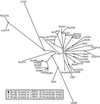
Myosin family tree. An unrooted phylogenetic tree of myosins shows the relationship of class III myosins to the overall myosin family. A clustal-W alignment (http://www2.ebi.ac.uk/clustalw/) was performed on the class III myosins known to date (Montell and Rubin, 1988; Hillman et al., 1996; Battelle et al., 1998; Wong et al., 1998; Doséet al., 2000; J. Lin-Jones [Gg], L. Sohlberg [Dr], personal communications) and one to three representative myosins from each of 18 classes. Kimura's correction was used and possibly led to the very long branches to DmIII and CeXII. The motor domains corresponding to residues 88–780 of chicken muscle myosin (Gg II) were compared using distance matrix analysis performed with clustal-W (http://bioweb.pasteur.fr/seqanal/interfaces/clustalw-simple.html), and the tree was drawn using TREEVIEW (http://taxonomy. zoology.gla.ac.uk/rod/treeview. html), which was further manipulated in Adobe Illustrator. Ac, Acanthamoeba casellanii; Acl, Acetabularia cliftonii; At, Aridopsis thaliana; Bt, Bos taurus; Ce, Caenorhabditis elegans; Dd, Dictyostelium discoidium; Dm, Drosophila melanogaster; Dr, Danio rerio; En, Emiricella nidulans; Gg, Gallus gallus; Ha, Helianthis annus; Hs, Homo sapiens; Lp, Liparis polyphemus; Mg, Magnaporthe grisea; Mm, Mus musculus; Ms, Morone saxatilis; Pf, Plasmodium falciparum; Rn, Rattus norvegicus; Tg, Toxoplasma gondii; Zm, Zea maize; nm, nonmuscle. All accession numbers are available on the Myosin Home Page (http://www.mrc-lmb.cam.ac.uk/myosin/trees/accession.html) except Hs MYO3A accession number NM017433, Hs MYO3B accession number NM138995, and Dr Myo3A accession number AF384863.
Figure 3
Figure 3
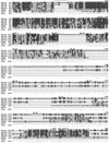
Amino acid sequence alignment of class III myosins. Boxshade output of a clustal W alignment (Young, 1976) of fish MyoIIIA (designated FMIIIA) and Myo3B (FMIIIB), human MYO3A (HMIIIA), human MYO3B (HMIIIB), Drosophila NINAC (DMIII), and Limulus MyoIIILim (LMIII). Black boxes indicate amino acid identity, and gray boxes indicate conserved amino acids. A dot (.) below the alignment indicates conserved amino acids, and an asterisk (*) indicates amino acids that are identical throughout all six class III myosins. The amino acid numbering for the individual sequences are at the left, and the boldfaced numbers above the sequences are for the overall alignment. The junctions between the kinase, head, neck, and tail domains are indicated. Overscored are the ATP-binding site within the kinase (ATP binding); the STE20 family signature motif (SSM); the two conserved ATP-binding regions at the myosin nucleotide binding cleft (NBC-1, or P-loop, and NBC-2); the region implicated in actin binding. The IQ motifs are italicized, and the two vertebrate class III myosin tail homology domains (3THDI and 3THDII) are indicated. Amino acid insertions are indicated (∧∧∧) above the sequence.
Figure 3
Figure 3
Amino acid sequence alignment of class III myosins. Boxshade output of a clustal W alignment (Young, 1976) of fish MyoIIIA (designated FMIIIA) and Myo3B (FMIIIB), human MYO3A (HMIIIA), human MYO3B (HMIIIB), Drosophila NINAC (DMIII), and Limulus MyoIIILim (LMIII). Black boxes indicate amino acid identity, and gray boxes indicate conserved amino acids. A dot (.) below the alignment indicates conserved amino acids, and an asterisk (*) indicates amino acids that are identical throughout all six class III myosins. The amino acid numbering for the individual sequences are at the left, and the boldfaced numbers above the sequences are for the overall alignment. The junctions between the kinase, head, neck, and tail domains are indicated. Overscored are the ATP-binding site within the kinase (ATP binding); the STE20 family signature motif (SSM); the two conserved ATP-binding regions at the myosin nucleotide binding cleft (NBC-1, or P-loop, and NBC-2); the region implicated in actin binding. The IQ motifs are italicized, and the two vertebrate class III myosin tail homology domains (3THDI and 3THDII) are indicated. Amino acid insertions are indicated (∧∧∧) above the sequence.
Figure 4
Figure 4
Expression patterns of Myo3A and Myo3B. Two DNA probes were generated for Northern blots: 3A, motor/tail, corresponding to amino acids 934-1203 of bass Myo3A, and 3B, kinase/motor, corresponding to amino acids 243–407 of bass Myo3B; these regions are designated on the bar diagrams in Figure 1. (B) Northern blot: 1.7 μg/lane of dark-adapted retina and RPE mRNA (left) and 4 μg/lane of light-adapted retina/RPE, brain, heart, intestine, kidney, liver, muscle, and testis (right) were probed with Myo3A- and Myo3B-specific probes. Myo3A is expressed at high levels in the retina and at lower levels in the brain and testis. Myo3B is expressed at more similar levels in the retina, intestine, and testis. Despite evidence of Myo3A expression in RPE by Western blot and immunohistochemistry, the Myo3A transcript is below detection levels on our Northern blot.
Figure 5
Figure 5
Western blot of Myo3A to determine relative protein levels in the retina and RPE and sacculus. Retina and RPE were dissected separately from dark-adapted bass as well as the sacculus: 9.4 μg of homogenized RPE (lane 1), retina (lane 2), and photoreceptor inner/outer segments (shake-offs) (lane 3) were loaded, and for comparison, 0.02 μg of Myo3A tail fusion protein (lane 4) was included. Also included are ∼10 μg of homogenized sacculus (lane 5). An antibody raised against a Myo3A tail-fusion protein also labels the ∼200-kDa band (α-tfp, lane 6). The RPE contains significantly less Myo3A than the retina, as does the sacculus (∼1/10th as determined by NIH Image), and the Myo3A is enriched in the sample containing photoreceptor inner/outer segments (shake-offs, greater than twofold increase in amount of Myo3A).
Figure 6
Figure 6
Myo3A immunolabeling of bass retinal tissue sections. Cryosections (8–10 μm) of dark-adapted bass retina were fixed in methanol and immunolabeled. Shown here are the Myo3A labeling (a), Myo3A labeling in the presence of 100-fold molar excess tail-tip peptide (b), and the DIC image (c). Label is concentrated in the distal ellipsoids of both rod and cone photoreceptors, and the specificity of the Myo3A antibody is confirmed by the elimination of epitope recognition in the presence of excess peptide. Cone ellipsoids (ce) and rod ellipsoids are designated by arrows. PL, photoreceptor layer; OLM, outer limiting membrane; ONL, outer nuclear layer. Scale bar, 10 μm.
Figure 7
Figure 7
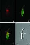
Colocalization of Myo3A and actin in the distal ellipsoid microfilament bundles and CPs of cone photoreceptors. Isolated cone photoreceptor inner/outer segments from dark-adapted bass retinas fixed in 4% paraformaldehyde were double-labeled with anti-Myo3A (a, red) and actin-labeling Alexa 488-phalloidin (b, green). The filamentous nature of the Myo3A staining coincides with the actin filament bundles of the inner segment and CPs at the outer-segment base, and the colocalization is seen as yellow in image overlays (c); (d) DIC image. The white arrows indicate the inner-segment/outer-segment junction. Scale bar, 10 μm.
Figure 8
Figure 8
Isolated rod photoreceptors double-labeled with Myo3A and anti-tubulin antibody. RIS/ROS were isolated from dark-adapted green sunfish retinas, fixed in methanol, and double-labeled with the Myo3A antibody (a, red), and anti-tubulin (a, green). An overlay with the DIC image (b, red) demonstrates that the Myo3A antibody labels the inner segment distal to the microtubules, suggesting that it is labeling primarily CPs. Scale bar, 2 μm.
Figure 9
Figure 9
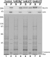
Triton X-100 extractions of RIS/ROS in the presence and absence of ATP. Top (Myo3A): In the absence of ATP (control) and in the presence of Mg2+ (1 mM Mg2+), all of the Myo3A associated with the pelleted cytoskeleton (P) and Myo3A was essentially absent from the Triton-solubilized fraction (S). In the presence of 1 mM ATP, some of the Myo3A was released from the cytoskeleton, and in the presence of 10 mM ATP, most of the Myo3A partitions with the supernatant, having been released from the cytoskeleton. Middle (actin): An anti-actin antibody demonstrates that in all cases, actin pellets with the Triton-insoluble fraction (P). Lower (total protein): A Coomassie blue stain shows that the majority of proteins are solubilized in Triton X-100 (S). Only the cytoskeleton and associated proteins are present in the pellet (P).
Figure 10
Figure 10
Electron micrographs of rod photoreceptor inner/outer-segment cytoskeletons. The detergent-insoluble fraction of isolated RIS/ROS was isolated and visualized by negative staining. Immunolabeling with an anti-Myo3A antibody abundantly decorates the actin filament bundles, indicating the presence of Myo3A along these filaments (A). The addition of ATP significantly reduces the Myo3A labeling, indicating the release of Myo3A from the actin filament bundles (B). The negative control with no primary shows little labeling (C). Scale bar, 1 μm.
Figure 11
Figure 11
Calmodulin affinity purification of bass Myo3A. The soluble protein fraction of light-adapted retina/RPE homogenate (lane 1) was tested for its ability to bind calmodulin. Like other myosins, bass Myo3A binds to calmodulin in the absence of calcium (lane 3). Having 17 μM calcium present during the incubation of calmodulin-Sepharose beads with retina/RPE extract may reduce the amount of Myo3A that binds to the calmodulin-Sepharose, but in general, the binding seems rather insensitive to calcium (lane 5). Under both conditions, a fraction of Myo3A does not bind calmodulin and remains in solution (lanes 2 and 4). When incubated with Sepharose beads alone, the majority of Myo3A remains in solution (lanes 6 and 7).

References

    1. Anderson DH, Johnson LV, Hageman GS. Vitronectin receptor expression and distribution at the photoreceptor-retinal pigment epithelial interface. J Comp Neurol. 1995;360:1–16. - PubMed
    1. Apweiler R, et al. Interpro—an integrated documentation resource for protein families, domains, and functional sites. Bioinformatics. 2000;16:1145–1150. - PubMed
    1. Arikawa K, Williams DS. Alpha-actinin and actin in the outer retina: a double immunoelectron microscopic study. Cell Motil Cytoskeleton. 1991;18:15–25. - PubMed
    1. Barnes JA, Gomes AV. PEST sequences in calmodulin-binding proteins. Mol Cell Biochem. 1995;149–150:17–27. - PubMed
    1. Barylko B, Binns DD, Albanesi JP. Regulation of the enzymatic and motor activities of myosin I. Biochim Biophys Acta. 2000;1496:23–35. - PubMed

Publication types

MeSH terms

Associated data

LinkOut - more resources