. 2003 Mar;14(3):1074-84.
doi: 10.1091/mbc.e02-05-0308.

Dynamin participates in focal extracellular matrix degradation by invasive cells

Affiliations

Dynamin participates in focal extracellular matrix degradation by invasive cells

Massimiliano Baldassarre et al. Mol Biol Cell. 2003 Mar.

Abstract

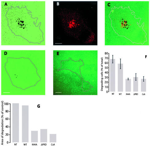
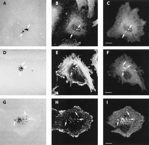
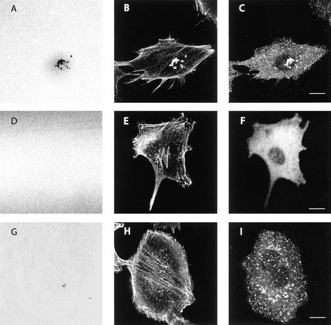
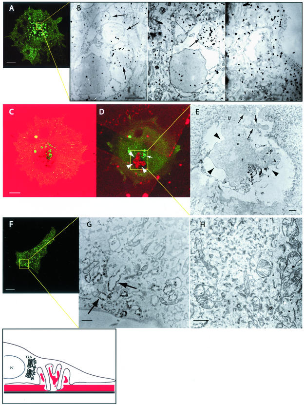
The degradation of extracellular matrix (ECM) by matrix metalloproteases is crucial in physiological and pathological cell invasion alike. Degradation occurs at specific sites where invasive cells make contact with the ECM via specialized plasma membrane protrusions termed invadopodia. Herein, we show that the dynamin 2 (Dyn2), a GTPase implicated in the control of actin-driven cytoskeletal remodeling events and membrane transport, is necessary for focalized matrix degradation at invadopodia. Dynamin was inhibited by using two approaches: 1) expression of dominant negative GTPase-impaired or proline-rich domain-deleted Dyn2 mutants; and 2) inhibition of the dynamin regulator calcineurin by cyclosporin A. In both cases, the number and extension of ECM degradation foci were drastically reduced. To understand the site and mechanism of dynamin action, the cellular structures devoted to ECM degradation were analyzed by correlative confocal light-electron microscopy. Invadopodia were found to be organized into a previously undescribed ECM-degradation structure consisting of a large invagination of the ventral plasma membrane surface in close spatial relationship with the Golgi complex. Dyn2 seemed to be concentrated at invadopodia.


Figures

Figure 1
Figure 1
ECM degradation by A375MM cells and effects of dominant negative dynamin 2 mutants and cyclosporin A on ECM degradation. A375MM melanoma cells were plated on cross-linked fluorophore-conjugated matrix for 15 h, fixed, stained with antigiantin antibodies to label the Golgi complex, and analyzed at the confocal microscope. A substrate level optical section shows the dark areas of degradation (A), the Golgi complex (B), and their relative positions in the merge (C). A375MM cells transiently transfected with wtDyn2aa-GFP, Dyn2(aa)K44A-GFP (D), or Dyn2(aa)ΔPRD-GFP (E) were cultured as described above and fixed. The percentage of degrading cells (F) and the areas of degradation (G) in nontransfected (NT), wild-type-, and mutant-transfected samples were quantified as described in Experimental Procedures Cell contours have been digitally rendered in the micrographs. Bar, 10 μm.
Figure 2
Figure 2
Localization of dynamin 2 and invadopodia markers to EDS. A375MM melanoma cells were plated on cross-linked fluorophore-conjugated matrix for 15 h, and then fixed, stained with anti-dynamin (Dyn2-ab), anti-cortactin, or anti-phosphotyrosine antibodies or phalloidin-TRITC and analyzed at the confocal microscope. ECM degradation patches (A, D, and G, arrows) from substrate level optical sections clearly coincided with either cortactin (B), actin (E), or phosphotyrosine (H) and a reinforcement of the Dyn2 signal (C, F, and I). Bar, 10 μm.
Figure 3
Figure 3
Effects of dominant negative dynamin 2 mutants on EDS. A375MM cells transiently transfected with wtDyn2aa-GFP (A–C), Dyn2(aa)ΔPRD-GFP (D–F), or Dyn2(aa)K44A-GFP (G–I) were cultured as described above, fixed, stained with anti-phalloidin-TRITC, and analyzed at the confocal microscope. Substrate level optical sections show the fluorescent matrix (A, D, and G), actin staining (B, E, and H), and the GFP signal (C, F, and I). Bar, 10 μm.
Figure 4
Figure 4
Correlative light-electron microscopy of EDS. Transfected A375MM melanoma cells were plated on CELLocate coverslips coated with cross-linked fluorophore-conjugated matrix for 15 h, fixed, analyzed at the confocal microscope, and then processed for immunoelectron microscopy. The shown electron microscopy fields are boxed in yellow in the corresponding confocal images, arrows indicate invadopodial protrusions. The boxed area of a wtDyn2aa-GFP–transfected cell (A) was serially sectioned and observed at the EM. The movie serial.mov shows the complete serial section series. Three different serial sections (bottom, middle, and top) (B) show Dyn2-GFP labeling as identified with anti-GFP antibodies. The movie stack.mov shows the complete Z-stack rendering of the confocal image series, whereas C and D show two different confocal sections of a same cell (substrate level and middle section, respectively); fragments of partially degraded gelatin are clearly visible both in the confocal (D) and electron microscopy images (E, arrowheads). The boxed area of Dyn2K44A-GFP–transfected cells (F) was observed at the electron microscope; arrows point to structures vaguely resembling EDS but devoid of invadopodia and gelatin fragments. H shows an electron micrograph of a Dyn2ΔPRD-GFP–transfected cell. The drawing depicts a schematic reconstruction of EDS in relationship to the nucleus (N) and the Golgi apparatus (GA), ECM is indicated in red. Bar, 10 μm in confocal images, and 1 μm in EM images.
Figure 5
Figure 5
Dynamics of invadopodia. A375MM melanoma cells, transiently transfected with wtDyn2aa-GFP, were plated on cross-linked fluorophore-conjugated matrix for 15 h and then analyzed at the confocal microscope at 37°C. (A) Digital capture of the cell imaged in the experiment shown: a single EDS (boxed area) was bleached for 30 s and the fluorescence allowed to recover for 80 s at 0.5 frames/s. FRAP is plotted as percentage of FRAP in time (B).

References

    1. Achiriloaie M, Barylko B, Albanesi JP. Essential role of the dynamin pleckstrin homology domain in receptor-mediated endocytosis. Mol Cell Biol. 1999;19:1410–1415. - PMC - PubMed
    1. Artalejo CR, Elhamdani A, Palfrey HC. Calmodulin is the divalent cation receptor for rapid endocytosis, but not exocytosis, in adrenal chromaffin cells. Neuron. 1996;16:195–205. - PubMed
    1. Awumey EM, Moonga BS, Sodam BR, Koval AP, Adebanjo OA, Kumegawa M, Zaidi M, Epstein S. Molecular and functional evidence for calcineurin-A α and β isoforms in the osteoclast: novel insights into cyclosporin A action on bone resorption. Biochem Biophys Res Commun. 1999;254:248–252. - PubMed
    1. Ayala I, Babia T, Baldassarre M, Pompeo A, Fabra A, Kok JW, Luini A, Buccione R, Egea G. Morphological and biochemical analysis of the secretory pathway in melanoma cells with distinct metastatic potential. FEBS Lett. 1999;451:315–320. - PubMed
    1. Backer JM. Phosphoinositide 3-kinases and the regulation of vesicular trafficking. Mol Cell Biol Res Commun. 2000;3:193–204. - PubMed

Publication types

MeSH terms

LinkOut - more resources