. 2003 Mar;14(3):1125-37.
doi: 10.1091/mbc.e02-08-0511.

Pre-M phase-promoting factor associates with annulate lamellae in Xenopus oocytes and egg extracts

Affiliations

Pre-M phase-promoting factor associates with annulate lamellae in Xenopus oocytes and egg extracts

Clare Beckhelling et al. Mol Biol Cell. 2003 Mar.

Abstract

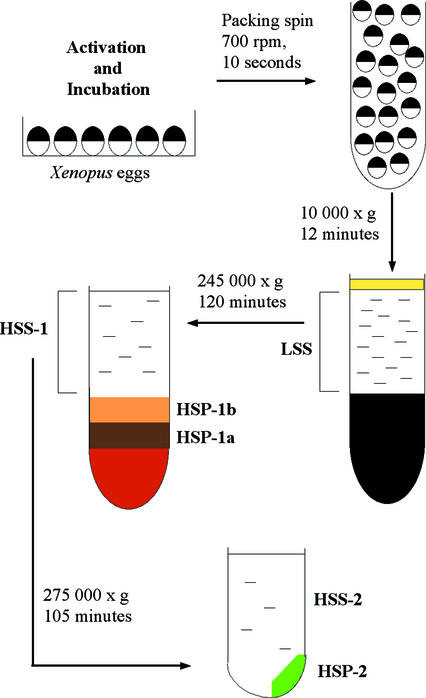
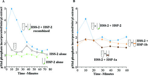
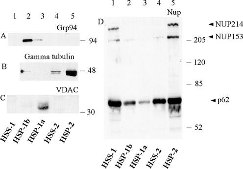
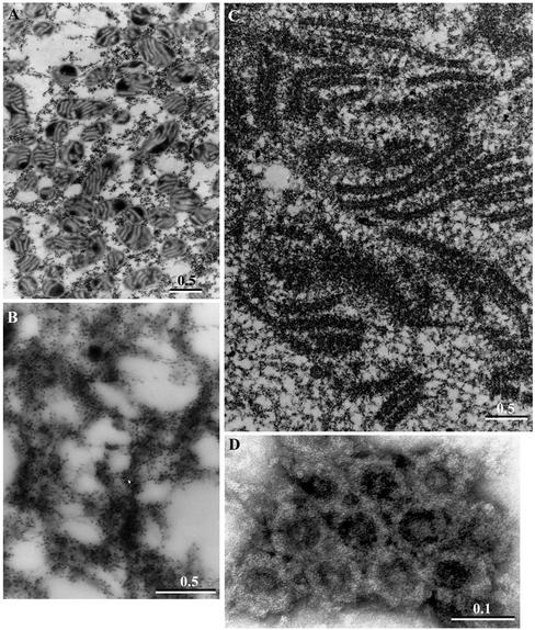
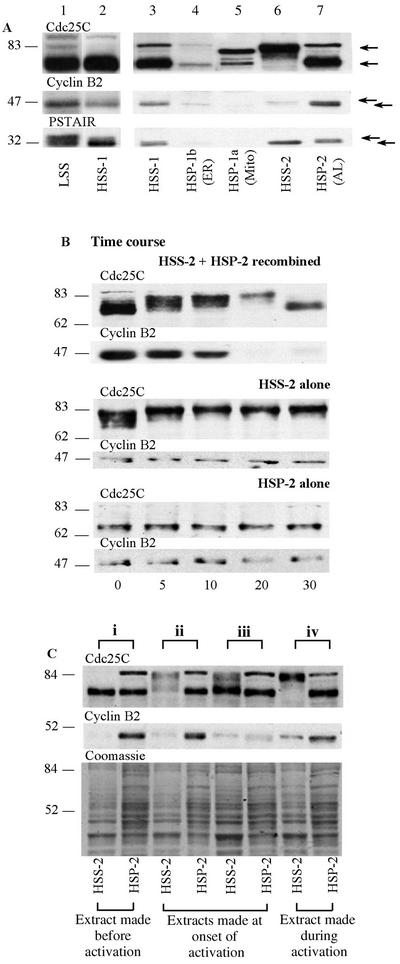
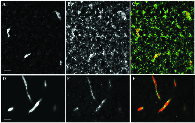
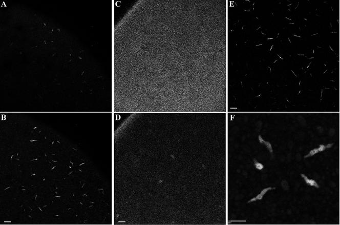
We have used complementary biochemical and in vivo approaches to study the compartmentalization of M phase-promoting factor (MPF) in prophase Xenopus eggs and oocytes. We first examined the distribution of MPF (Cdc2/CyclinB2) and membranous organelles in high-speed extracts of Xenopus eggs made during mitotic prophase. These extracts were found to lack mitochondria, Golgi membranes, and most endoplasmic reticulum (ER) but to contain the bulk of the pre-MPF pool. This pre-MPF could be pelleted by further centrifugation along with components necessary to activate it. On activation, Cdc2/CyclinB2 moved into the soluble fraction. Electron microscopy and Western blot analysis showed that the pre-MPF pellet contained a specific ER subdomain comprising "annulate lamellae" (AL): stacked ER membranes highly enriched in nuclear pores. Colocalization of pre-MPF with AL was demonstrated by anti-CyclinB2 immunofluorescence in prophase oocytes, in which AL are positioned close to the vegetal surface. Green fluorescent protein-CyclinB2 expressed in oocytes also localized at AL. These data suggest that inactive MPF associates with nuclear envelope components just before activation. This association may explain why nuclei and centrosomes stimulate MPF activation and provide a mechanism for targeting of MPF to some of its key substrates.


Figures

Figure 1
Figure 1
Preparation of high-speed fractions of egg extracts. Eggs, taken 60 min after activation, were subject to a brief packing spin before low-speed centrifugation to crush them, separating supernatant (LSS) from yolk pellet and lipid layer. High-speed centrifugation stratified supernatant (HSS-1) from differently colored pellet fractions (HSP-1a, HSP-1b) above a gelatinous orange layer that was discarded. Dilution and further centrifugation of HSS-1 generated HSS-2 and HSP-2. See MATERIALS AND METHODS for detailed explanation.
Figure 2
Figure 2
Histone (H1) kinase activity of fractions incubated separately and in combination. Samples of each fraction were either diluted into extraction buffer or combined with HSS-2 before transfer to 21°C. Aliquots were taken every 10 min for up to 80 min for assay of histone (H1) kinase activity, expressed as picomoles of phosphate transferred per minute per microliter of extract. (A) HSS-2 alone (open squares), HSP-2 alone (green triangles), and HSS-2 recombined with HSP-2 (blue circles). Neither fraction can activate H1 kinase alone but when recombined stimulate one cycle of activation and inactivation of H1 kinase. (B) From a different extract, HSS-2 was recombined with either HSP-2 (blue circles), HSP-1a (brown circles), or HSP-1b (yellow triangles). Only HSP-2 stimulated one cycle of mitotic histone (H1) kinase activation and inactivation when combined with HSS-2.
Figure 3
Figure 3
Enrichment of nuclear pore proteins in HSP-2. Twenty micrograms (as determined by Bradford assay) of each fraction obtained by serial centrifugation of prophase extracts (Figure 1) was loaded onto different percentages of gels depending upon the antibody to be used, transferred to nitrocellulose, and probed with the antibodies indicated. HSP-1b was highly enriched in ER protein, GRP94 (A, lane 2), whereas HSP-1a was enriched in the mitochondrial protein VDAC (C, lane 3). HSP-2 contained three nuclear pore proteins detected by the anti-NUP antibody: NUP214, NUP153, and p62 (arrowheads in D, lane 5). HSP-2 was also enriched in γ-tubulin (B, lane 5). In all immunoblots, lane 1 contains an unfractionated HSS-1 and lane 4 contains the HSS-2 made by dilution and recentrifugation of HSS-1.
Figure 4
Figure 4
Annulate lamellae are enriched in HSP-2. TEM and negative staining were used to identify structures in the pelleted fractions. (A) Image of HSP-1a, showing mitochondria and some putative ribosomal material. (B) Image of HSP-1b, membranes covered in ribosome-sized particles consistent with an enrichment of ER. (C) Image of HSP-2, containing granular material and stacks of membranes containing identifiable nuclear pore complexes. The membranes have been sectioned both transversely and tangentially. Pore complexes were also detected by negative staining in HSP-2 from another extract (D). Bars, 0.5 μm in A–C and 0.1 μm in D.
Figure 5
Figure 5
(A) Pre-MPF and inactive Cdc25C preferentially pellet into HSP-2. Immunoblots showing the different isoforms of MPF and Cdc25C in prophase LSS and HSS-1 run on the same gel (lanes 1 and 2) and in the various high-speed fractions run on a second gel (lanes 3–7, equal loading was confirmed by Coomassie blue staining, our unpublished data). After electrophoretic transfer, different molecular weight regions of the same blot were probed with the antibodies indicated above each horizontal strip. Most of the CyclinB2 and Cdc2 present in the initial LSS extract remained in the supernatant (HSS-1) after high-speed centrifugation. The second high-speed centrifugation pelleted most inactive Cdc25C (lower arrow, hypophosphorylated form) and inactive, phosphorylated Cdc2 (upper arrow in PSTAIR blot), together with most of the CyclinB2, representing the pre-MPF predominant in these extracts (lane 7). In contrast, the small amount of active MPF present in the original prophase extracts was retained in the final soluble fraction (HSS-2, lane 6), as indicated by the presence of CyclinB2 predominantly in the phosphorylated form (upper arrow) associated with activating/activated MPF and the absence of the inactive Cdc2 isoforms. Multiply phosphorylated Cdc25C (upper arrow) was also present in the HSS-2. (B) Cdc25C can activate in the supernatant alone but not in the pellet alone. Volumes (30 μl) of either recombined HSS-2 + HSP-2 or HSS-2 alone or HSP-2 alone were transferred to 21°C and aliquots were taken every 5–10 min for 30 min (indicated at the bottom of the figure). A typical time course of activation (phosphorylation and shift to the upper isoforms) and inactivation (dephosphorylation and down shift on the gel) of Cdc25C occurred >30 min in the recombined aliquots (top). Activation without inactivation was observed in the HSS-2 alone, whereas no activation occurred in the HSP-2 alone. Approximate relative molecular weights in kilodaltons are shown on the left. (C) Activated forms of CyclinB2 shift from HSP-2 to HSS-2 during MPF activation. The activation status of Cdc25C and presence of CyclinB2 in HSS-2 and HSP-2 were compared in four different extracts made at slightly different times in the cell cycle and designated i to iv as indicated at the top of the figure. From analysis of MPF activation in these extracts at 21°C (see text for details), we can deduce that extract i was made before activation, extracts ii and iii were made at the onset of activation, and extract iv was made during the activation of MPF, as stated at the bottom of the figure. The same region of a parallel gel stained with Coomassie blue is shown to allow comparison of protein loading. Approximate relative molecular weights in kilodaltons are shown on the left. For each extract i to iv paired samples of HSS-2 (left) and HSP-2 (right) were loaded as indicated below the lanes. Irrespective of the degree of MPF activation in these extracts, most of the faster migrating CyclinB2 isoform, representing pre-MPF (A) and inactive Cdc25C is pelleted into HSP-2.
Figure 6
Figure 6
Detection of CyclinB2 at AL in stage VI oocytes. Confocal images of the vegetal cortex region of oocytes fixed and processed for double immunofluorescence by using anti-Nup and anti-GRP94 (A and B) and anti-Nup and anti-CyclinB2 (D and E). Superimposed images with anti-GRP94 or anti-CyclinB2 in green and anti-NUP in red are shown in C and F, respectively. Bars, 10 μm.
Figure 7
Figure 7
GFP-CyclinB2 localization to annulate lamellae in vivo. (A and B) An oocyte microinjected with CyclinB2-GFP mRNA and imaged by confocal microscopy 4 h later from the vegetal side. A shows the vegetal cortical layer, whereas B is at the level of the subcortical AL. (C and D) An oocyte microinjected with nonconjugated GFP mRNA. C shows the vegetal cortex, whereas D is a subcortical layer equivalent to that in B. Note in D the fluorescence in the oocyte cortical rim that is not apparent with the GFP-CyclinB2 in B. E a confocal image of AL labeled by immunofluorescence in a fixed oocyte by the anti-NUP antibody at the same magnification as A–D. Bars, 20 μm. F is a higher magnification image of islands labeled in B by the GFP-CyclinB2. Bar, 10 μm.

Similar articles

Cited by

References

    1. Alfa CE, Ducommun B, Beach D, Hyams JS. Distinct nuclear and spindle pole body population of cyclin-cdc2 in fission yeast. Nature. 1990;347:680–682. - PubMed
    1. Argon Y, Simen BB. GRP94, an ER chaperone with protein and peptide binding properties. Semin Cell Dev Biol. 1999;10:495–505. - PubMed
    1. Ashcroft NR, Srayko M, Kosinski ME, Mains PE, Golden A. RNA-Mediated interference of a cdc25 homolog in Caenorhabditis elegans results in defects in the embryonic cortical membrane, meiosis, and mitosis. Dev Biol. 1999;206:15–32. - PubMed
    1. Audit M, Barbier M, Soyer-Gobillard MO, Albert M, Géraud ML, Nicolas G, Lenaers G. CyclinB (p56cdc13) localization in yeast Schizosaccharomyces pombe: an ultrastructural and immunocytochemical study. Biol Cell. 1996;86:1–10. - PubMed
    1. Bagley S, Goldberg MW, Cronshaw JM, Rutherford S, Allen TD. The nuclear pore complex. J Cell Sci. 2000;113:3885–3886. - PubMed

Publication types

MeSH terms

LinkOut - more resources