. 2003 Mar;14(3):1138-48.
doi: 10.1091/mbc.e02-07-0411.

Obscurin is a ligand for small ankyrin 1 in skeletal muscle

Affiliations

Obscurin is a ligand for small ankyrin 1 in skeletal muscle

Aikaterini Kontrogianni-Konstantopoulos et al. Mol Biol Cell. 2003 Mar.

Abstract

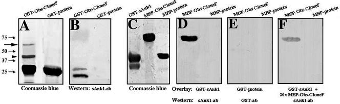
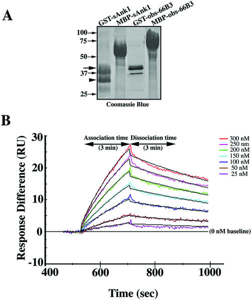
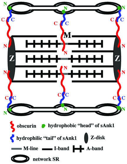
The factors that organize the internal membranes of cells are still poorly understood. We have been addressing this question using striated muscle cells, which have regular arrays of membranes that associate with the contractile apparatus in stereotypic patterns. Here we examine links between contractile structures and the sarcoplasmic reticulum (SR) established by small ankyrin 1 (sAnk1), a approximately 17.5-kDa integral protein of network SR. We used yeast two-hybrid to identify obscurin, a giant Rho-GEF protein, as the major cytoplasmic ligand for sAnk1. The binding of obscurin to the cytoplasmic sequence of sAnk1 is mediated by a sequence of obscurin that is C-terminal to its last Ig-like domain. Binding was confirmed in two in vitro assays. In one, GST-obscurin, bound to glutathione-matrix, specifically adsorbed native sAnk1 from muscle homogenates. In the second, MBP-obscurin bound recombinant GST-sAnk1 in nitrocellulose blots. Kinetic studies using surface plasmon resonance yielded a K(D) = 130 nM. On subcellular fractionation, obscurin was concentrated in the myofibrillar fraction, consistent with its identification as sarcomeric protein. Nevertheless, obscurin, like sAnk1, concentrated around Z-disks and M-lines of striated muscle. Our findings suggest that obscurin binds sAnk1, and are the first to document a specific and direct interaction between proteins of the sarcomere and the SR.


Figures

Figure 1
Figure 1
Yeast two-hybrid analysis identified obscurin as the major cytoplasmic ligand for sAnk1. (A) The COOH-terminal hydrophilic tail of sAnk129–155 was used as bait to screen a rat skeletal muscle cDNA library. (B) Six of the 12 positive preys encoded overlapping fragments of the 2.7-kb, COOH-terminal sequence of obscurin.
Figure 2
Figure 2
Sequence of the C-terminal region of rat obscurin. (A) Sequence comparison between the COOH-terminal sequence of the human and rat obscurin orthologues. The first line represents amino acids5816–6620 of the human protein (Young et al., 2001), the third line shows the corresponding rat sequence, and the middle line shows shared residues. Overall homology and similarity are ∼77% and ∼83%, respectively. The Ig-like, PH and (partial) Rho-GEF domains are in boxes. The positions of obscurin clones A–F are denoted by arrows. (B) A 525 base pairs 3′UTR is carried by all six obscurin clones; the putative polyadenylation signal is underlined.
Figure 3
Figure 3
Regions of sAnk1 and obscurin involved in binding. To identify the minimal sequences of sAnk1 and obscurin required for their association, a series of deletion constructs were generated and expressed in the pLexA-bait and pB42AD-prey vectors respectively. Yeast two-hybrid analysis followed by a β-galactosidase assay in solution indicated that these were confined to a 70 amino acid sequence in sAnk1 (sAnk1aa61–130) and a 120-amino acid segment in obscuring (obscurinaa501–627) that corresponds to amino acid sequences 6312–6432 of the human obscurin homologue.
Figure 4
Figure 4
Direct, specific binding of sAnk1 to obscurin in vitro. (A) Equivalent amounts of GST-obscurin clone F fusion protein and GST were subjected to SDS-PAGE and stained with Coomassie blue. The GST-obscurin clone F fusion protein yielded two products, the intact protein at ∼64 kDa (arrow) and a major breakdown product at ∼45 kDa (arrowhead). GST-protein is ∼25 kDa. (B) After binding to glutathione matrices, recombinant GST-obscurin clone F, but not GST, adsorbed native sAnk1 from homogenates of quadriceps muscle, as shown by SDS-PAGE and immunoblotting with antibodies to sAnk1. (C) Bacterially expressed GST-sAnk1 (∼39 kDa), MBP-obscurin clone F (∼80 kDa), and MBP-protein (∼42 kDa) were separated by SDS-PAGE and stained with Coomassie blue. (D) Purified GST-sAnk129–155 binds to immobilized MBP-obscurin clone F fusion protein but not to MBP, as shown by blotting with antibodies to sAnk1. (E) No binding to MBP-obscurin or MBP was detected in blots similar to those in D incubated with GST alone and probed with antibodies to GST. (F) The specific binding seen in D is significantly reduced by adding a 20-fold excess of MBP-obscurin clone F fusion protein to the overlay buffer along with recombinant GST-sAnk129–155.
Figure 5
Figure 5
Real time kinetic analysis of the binding of sAnk1 to obscurin by surface plasmon resonance. (A) Bacterially expressed, affinity purified GST-sAnk1 (∼39 kDa), MBP-sAnk1 (∼57 kDa), GST-obscurin-F3 (∼38.5 kDa), and MBP-obscurin-F3 (∼66 kDa) were fractionated on SDS-PAGE followed by Coomassie blue staining. Note the heterogeneity of the GST-sAnk1 construct, which contains a major breakdown product at ∼32 kDa (arrowhead) along with the intact protein (39 kDa, arrow). (B) A series of binding curves were generated in real time (experiment 1 of Table 1) in which GST-obscurin-F3 was used as ligand and MBP-sAnk1, at concentrations from 0 to 300 nM, as analyte. In this experiment, immobilized GST-obscurin-F3 was present in FC2, and GST in FC1. After correction for nonspecific binding and alignment of the sensograms (see RESULTS), curves of the data, shown in different colors, were fitted with the 1:1 Langmuir binding model, shown in black. The y-axis indicates the resonance units (RU) generated by the binding of MBP-sAnk1 to surface-bound GST-obscurin-F3, with each curve representing a different concentration of analyte. The results are consistent with 1:1 binding of the sAnk1 and obscurin fusion proteins, with an affinity of 130 nM (Table 1).
Figure 6
Figure 6
Obscurin is present in the detergent insoluble fraction of muscle lysates. (A) Western blotting with antibodies to obscurin clone B shows that obscurin is primarily in the detergent insoluble (DI) pellet of adult skeletal muscle lysates (arrow). Smaller amounts of obscurin are detected in the detergent soluble (DS) fractions. (B) Unlike obscurin, sAnk1 (arrowheads) is enriched in the DS fraction.
Figure 7
Figure 7
The subcellular distribution of obscurin in skeletal and cardiac myofibers. (A–C) Obscurin (A) is present in striations in longitudinal sections of quadriceps myofibers. Lines correspond to Z-disks (arrows) and M-lines (arrowheads), as shown by double immunostaining with antibodies to the Z-disk marker α-actinin (B). In the color overlay (C), obscurin is in red, α-actinin in green, and areas with both proteins in yellow. (D) Labeling for obscurin shows a reticular pattern in cross sections of quadriceps skeletal muscle. (E) Prominent staining of obscurin occurs at M-lines (arrowhead) in adult myocardium, with weaker labeling at Z-disks (arrow), marked by anti–α-actinin. Colors are as in panel C. (F) Like obscurin, sAnk1 in cardiac muscle concentrates over M-lines (arrowhead) and to a lesser extent around Z-disks (arrow), stained with anti–α-actinin. In (F), sAnk1 is shown in red and α-actinin in green. (G) No labeling was detected when nonimmune rabbit IgG was used. H. Preabsorption of the obscurin antibody with its antigen eliminated the labeling observed in panel A.
Figure 8
Figure 8
Model of sAnk1 association with obscurin. sAnk1 is anchored to the network SR, around Z-disks and M-lines, through its hydrophobic NH2-terminus, with its hydrophilic COOH-terminal portion facing the myoplasm. Obscurin appears to surround the myofibrils at Z-disks and M-lines, with its COOH-terminus exposed to allow it to bind to sAnk1. Thus, sAnk1 binding to obscurin may contribute to the sarcomeric alignment of the network SR.

Similar articles

Cited by

References

    1. Bang ML, et al. The complete gene sequence of titin, expression of an unusual approximately 700-kDa titin isoform, and its interaction with obscurin identify a novel Z-line to I-band linking system. Circ Res. 2001;89:1065–1072. - PubMed
    1. Baumann O, Walz B. Endoplasmic reticulum of animal cells and its organization into structural and functional domains. Int Rev Cytol. 2001;205:149–214. - PubMed
    1. Bennett V. Ankyrins. Adaptors between diverse plasma membrane proteins and the cytoplasm. J Biol Chem. 1992;267:8703–8706. - PubMed
    1. Bennett V, Chen L. Ankyrins and cellular targeting of diverse membrane proteins to physiological sites. Curr Opin Cell Biol. 2001;13:61–67. - PubMed
    1. Birkenmeier CS, White RA, Peters LL, Hall EJ, Lux SE, Barker JE. Complex patterns of sequence variation and multiple 5′ and 3′ ends are found among transcripts of the erythroid ankyrin gene. J Biol Chem. 1993;81:2144–2149. - PubMed

Publication types

MeSH terms

Associated data

LinkOut - more resources