. 2003 Mar;14(3):1182-94.
doi: 10.1091/mbc.e02-03-0167.

Essential roles for GPI-anchored proteins in African trypanosomes revealed using mutants deficient in GPI8

Affiliations

Essential roles for GPI-anchored proteins in African trypanosomes revealed using mutants deficient in GPI8

Simon Lillico et al. Mol Biol Cell. 2003 Mar.

Abstract

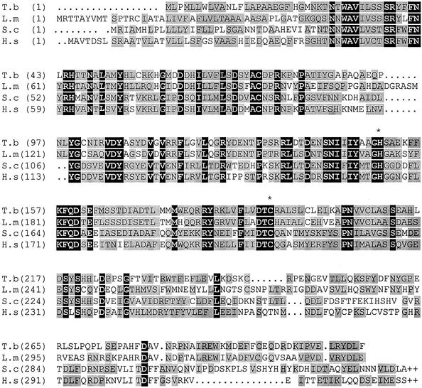
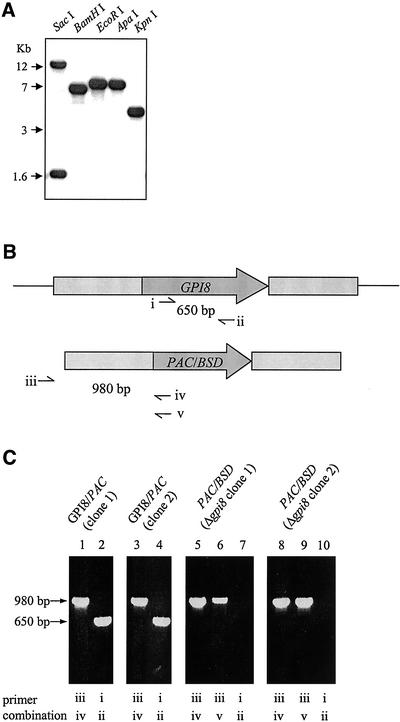
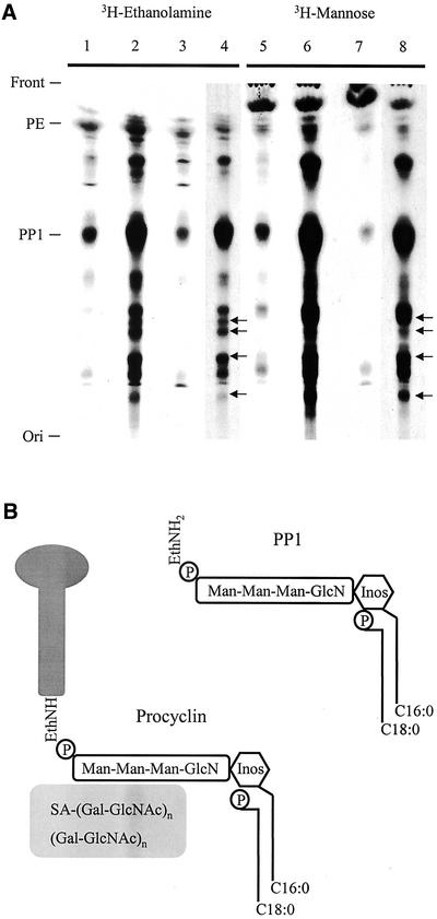
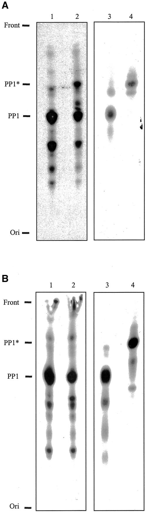
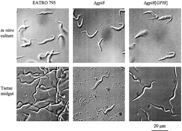
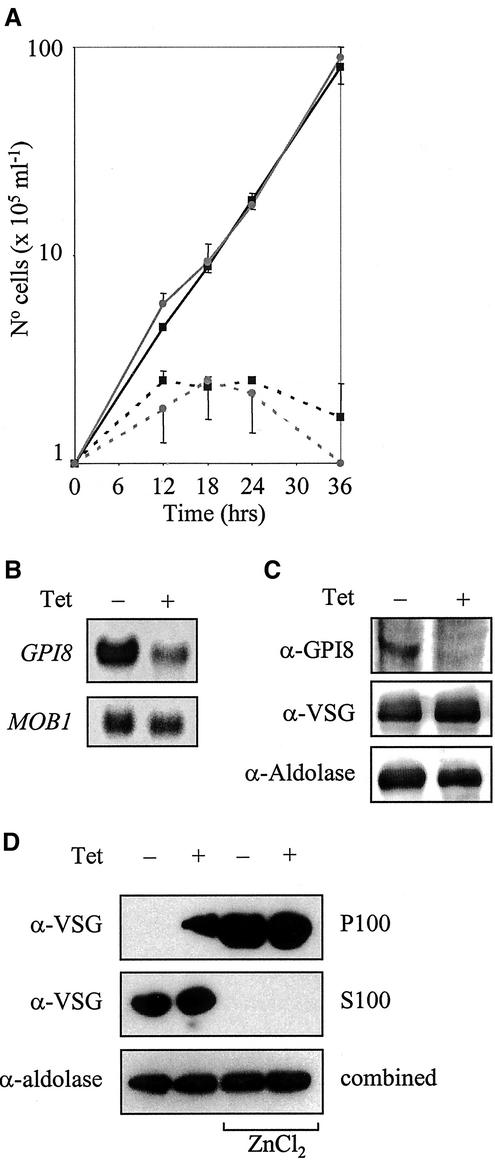
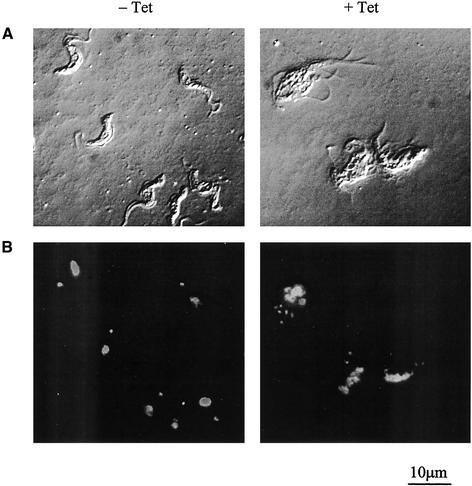
The survival of Trypanosoma brucei, the causative agent of Sleeping Sickness and Nagana, is facilitated by the expression of a dense surface coat of glycosylphosphatidylinositol (GPI)-anchored proteins in both its mammalian and tsetse fly hosts. We have characterized T. brucei GPI8, the gene encoding the catalytic subunit of the GPI:protein transamidase complex that adds preformed GPI anchors onto nascent polypeptides. Deletion of GPI8 (to give Deltagpi8) resulted in the absence of GPI-anchored proteins from the cell surface of procyclic form trypanosomes and accumulation of a pool of non-protein-linked GPI molecules, some of which are surface located. Procyclic Deltagpi8, while viable in culture, were unable to establish infections in the tsetse midgut, confirming that GPI-anchored proteins are essential for insect-parasite interactions. Applying specific inducible GPI8 RNAi with bloodstream form parasites resulted in accumulation of unanchored variant surface glycoprotein and cell death with a defined multinuclear, multikinetoplast, and multiflagellar phenotype indicative of a block in cytokinesis. These data show that GPI-anchored proteins are essential for the viability of bloodstream form trypanosomes even in the absence of immune challenge and imply that GPI8 is important for proper cell cycle progression.


Figures

Figure 1
Figure 1
Multiple alignment of GPI8 amino acid sequences from T. brucei (this study), Leishmania mexicana (CAB55340), S. cerevisiae (NP_010618) and Homo sapiens (CAA68871). The alignment was performed using Align X (Vector NTI Suite, Informax Inc.). Amino acids identical in all sequences are highlighted with a black background, and gray blocks denote conservation. Active site cysteine and histidine residues are indicated by an asterisk (∗).
Figure 2
Figure 2
Generation of Δgpi8 mutants (A) Southern blot analysis of the T. brucei GPI8 gene. DNA was cut with SacI, BamHI, EcoRI, ApaI, or KpnI, transferred onto nylon membrane and hybridized with the ORF of the gene. (B) Schematic representation of the GPI8 locus (top) and PAC/BSD replacement construct (bottom) with open reading frames (dark gray) and GPI8 flanking regions (light gray) used in the integration construct. Thin lines represent regions not present in the integration construct. Primer sites used to confirm presence of GPI8 (i and ii) and correct integration of PAC/BSD constructs (iii with iv or v) are indicated. (C) PCR of genomic DNA to determine correct integration of markers and deletion of GPI8. GPI8/PAC (clone 1) and (clone 2) are two independent heterozygotes containing a PAC (lanes 1 and 3) and a GPI8 (lanes 2 and 4) gene. Derived from these are two independent Δgpi8 clones, containing PAC (lanes 5 and 8) and BSD (lanes 6 and 9), but lacking GPI8 (lanes 7 and 10). Δgpi8 clone 1 was used for all subsequent studies. Some results were confirmed with clone 2 (unpublished data).
Figure 3
Figure 3
Analysis of procyclin expression (A) Western blot analysis of whole cell lysate from wild-type EATRO 795, Δgpi8 and Δgpi8[GPI8] detected with α-EP procyclin, α-GPEET procyclin, or α-aldolase. (B) Wild-type EATRO 795, Δgpi8, and Δgpi8[GPI8] were hypotonically lysed, and the washed pellet was subjected to CHAPS extraction. After SDS-PAGE, CHAPS extracted protein was detected with Stainsall or SyproRuby. Arrows indicate abundant proteins present in all three samples (←) or predominantly in the wild-type EATRO 795 and Δgpi8[GPI8] or Δgpi8 (→).
Figure 4
Figure 4
Accumulation of lipids in Δgpi8. (A) Chloroform/methanol/water extracts of 3H-ethanolamine- and 3H-mannose–labeled procyclic forms were chromatographed and visualized by autoradiography. Δgpi8[GPI8] (lanes 1 and 5), Δgpi8 (lanes 2, 4, 6, and 8), and wild-type EATRO 795 (lanes 3 and 7). The positions of the front, origin, phosphatidylethanolamine (PE), and the major insect-stage T. brucei GPI-anchor precursor PP1 (Field et al., 1991b) are indicated at left. A number of novel lipid species were detected in Δgpi8 (indicated by arrows [←]). For clarity, the Δgpi8 extracts are shown after both 2-d (lanes 4 and 8) and 10-d (lanes 2 and 6) exposures. (B) Schematic illustrating the structural features of PP1 and the procyclin GPI anchor. Data are based on studies cited in the text. PP1, top right, contains a cannonical GPI-core glycan, together with an acyl-inositol headgroup (the major fatty acid here is palmitate, although other species are present) and a lyso-glycerolipid backbone (a single fatty acid, predominantly stearate, is attached to the glycerol sn-1 position). The procyclin GPI-anchor structure has the same core as PP1, including an identical fatty acid configuration. Additionally, the procyclin protein C terminus is in amide linkage to the ethanolamine, and there is a large heterogenous glycan of ∼15-kDa molecular weight, which contains sialic acid and is endo-β-galactosidase sensitive (indicating the presence of lactosamine repeat structures). Inositol is represented by a hexagon, the core glycan, and the side chain in the procyclin anchor by rounded rectangles, and the procyclin polypeptide by a lollipop. Diagram is not to scale.
Figure 5
Figure 5
Surface expression of protein-anchor precursors. (A) Δgpi8 cells labeled with 3H-mannose were chased for 4 h in fresh medium then incubated without (lane 1) or with (lane 2) 10 mM sodium periodate. After quenching of the oxidation reaction, cell pellets were extracted sequentially with chloroform/methanol and chloroform/methanol/water, chromatographed, and visualized by autoradiography. The chloroform/methanol fraction demonstrated even loading of samples as determined by phosphatidylethanolamine abundance. The positions of the front, origin, PP1, and oxidized PP1 (PPI*) in the chloroform/methanol/water fraction are indicated on the left. To provide an authentic standard for PP1*, PP1 was purified by elution from a TLC plate and treated without (lane 3) and with (lane 4) 10 mM periodate for 10 min. Samples were chromatographed and visualized by autoradiography. (B) Δgpi8 cells labeled for 2.5 h with 3H-mannose and then incubated without (lane 1) or with (lane 2) 10 mM sodium periodate. Lipids extracted from Δgpi8 cells were incubated in the absence (lane 3) or presence (lane 4) of 10 mM periodate for 10 min. Samples were chromatographed as in A.
Figure 6
Figure 6
Tsetse fly infectivity. Wild-type EATRO 795, Δgpi8, and Δgpi8[GPI8] were fed to teneral tsetse flies as part of a blood meal. After 14 d, midguts were dissected and examined microscopically. Images of living parasites from in vitro cultures and tsetse dissections are displayed.
Figure 7
Figure 7
RNAi of bloodstream form trypanosomes. (A) Growth curve of two independently derived clones in the absence (solid line) or presence (dashed line) of 1 μg ml−1 tetracycline. The experiment was carried out in triplicate and bars denoting SD are shown. (B) Northern blots of total RNA prepared from parasites 12 h −/+ induction, electrophoresed on a 1% agarose gel and transferred onto nylon membrane. Duplicate blots were hybridized with 32P-labeled ORFs of either GPI8 or MOB1. (C) Western blot analysis of whole cell lysate 24 h −/+ induction probed with α-GPI8, α-VSG 221, or α-aldolase. A Typhoon 8600 phosphoimager was used for detection and quantitation. (D) Cells, 1 × 10⁷, cultured for 24 h −/+ induction were lysed on ice in 25 μl 0.05% TX-100 with 1 mg ml−1 pefabloc and 1 μg ml−1 leupeptin and then incubated at 37°C for 30 min. Alternatively cells were lysed as above in the presence of 10 mM ZnCl2 and maintained on ice for 5 min. Lysates were made up to 1 ml, after which they were centrifuged at 100,000 × g for 30 min at 4°C to yield P100 and S100 fractions. Samples were analyzed by Western blotting with α-VSG 221 or α-aldolase.
Figure 8
Figure 8
Microscopic analysis of cultured bloodstream form parasites without (−) or with (+) 18 h tetracycline induction of GPI8 RNAi using (A) phase contrast or (B) DAPI staining of DNA.

References

    1. Acosta-Serrano A, Cole RN, Englund PT. Killing of Trypanosoma brucei by concanavalin A: structural basis of resistance in glycosylation mutants. J Mol Biol. 2000;304:633–644. - PubMed
    1. Acosta-Serrano A, Vassella E, Liniger M, Renggli CK, Brun R, Roditi I, Englund PT. The surface coat of procyclic Trypanosoma brucei: programmed expression and proteolytic cleavage of procyclin in the tsetse fly. Proc Natl Acad Sci USA. 2001;98:1513–1518. - PMC - PubMed
    1. Bangs JD, Ransom DM, McDowell MA, Brouch EM. Expression of bloodstream variant surface glcoproteins in procyclic stage Trypanosoma brucei: role of GPI anchors in secretion. EMBO J. 1997;16:4285–4294. - PMC - PubMed
    1. Barrett AJ, Rawlings ND. Evolutionary lines of cysteine peptidases. Biol Chem. 2001;382:727–733. - PubMed
    1. Baumann NA, Vidugiriene J, Machamer CE, Menon AK. Cell surface display and intracellular trafficking of free glycosylphosphatidylinositols in mammalian cells. J Biol Chem. 2000;275:7378–7389. - PubMed

Publication types

MeSH terms

LinkOut - more resources