. 2003 Mar;14(3):1204-20.
doi: 10.1091/mbc.e02-09-0609.

Tor kinases are in distinct membrane-associated protein complexes in Saccharomyces cerevisiae

Affiliations

Tor kinases are in distinct membrane-associated protein complexes in Saccharomyces cerevisiae

Karen P Wedaman et al. Mol Biol Cell. 2003 Mar.

Abstract

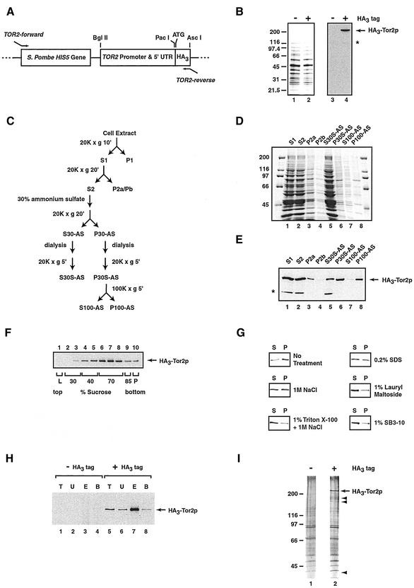
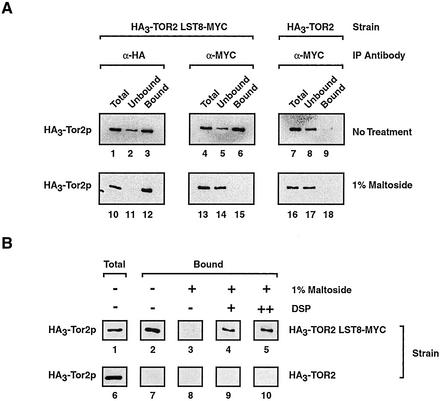
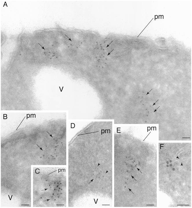
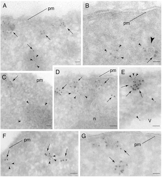
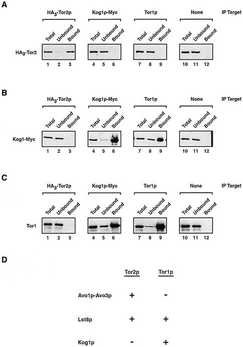
Tor1p and Tor2p kinases, targets of the immune-suppressive antibiotic rapamycin, are components of a highly conserved signaling network that couples nutrient availability and cell growth. To gain insight into the molecular basis underlying Tor-dependent signaling, we used cell fractionation and immunoaffinity chromatography to examine the physical environment of Tor2p. We found that the majority of Tor2p associates with a membrane-bound compartment along with at least four other proteins, Avo1p-Avo3p and Lst8p. Using immunogold electron microscopy, we observed that Tor2p, as well as Tor1p, localizes in punctate clusters to regions adjacent to the plasma membrane and within the cell interior, often in association with characteristic membranous tracks. Cell fractionation, coimmunoprecipitation, and immunogold electron microscopy experiments confirmed that Lst8 associates with both Tor2p as well as Tor1p at these membranous sites. In contrast, we find that Kog1, the yeast homologue of the mammalian Tor regulatory protein Raptor, interacts preferentially with Tor1p. These findings provide evidence for the existence of Tor signaling complexes that contain distinct as well as overlapping components. That these complexes colocalize to a membrane-bound compartment suggests an intimate relationship between membrane-mediated signaling and Tor activity.


Figures

Figure 1
Figure 1
Strategy used to isolate Tor2p copurifying proteins. (A) Relevant portion of plasmid pFA6a-HisMX6-PTOR2-ATG-3HA, a derivative of pFA6a-His3MX6-PGAL1–3HA (Longtine et al., 1998), used to construct strain HA3-TOR2, as described in MATERIALS AND METHODS. Indicated are relevant restriction endonuclease sites, the translational start site, and primers used for PCR-mediated integration of the diagrammed cassette. (B) Characterization of HA3-Tor2. Western blot analysis of extracts prepared from HA3-TOR2 or W303a, indicated by + or −-HA3 tag, respectively (right). For comparison, a Coomassie-stained gel of each extract is also shown (left). (C) Scheme used to prepare HA3-Tor2p–enriched fraction P100-AS. Indicated are the relevant ultracentrifugation, and ammonium sulfate precipitation and dialysis steps, as described in detail in the text. (D) Coomassie-stained gel of total proteins in fractions, corresponding to steps outlined in C prepared during preparation of P100-AS. (E) Western blot analysis detecting HA3-Tor2p in fractions corresponding to those in D. (F) Equilibrium density centrifugation of the P100-AS fraction on a sucrose step gradient. After centrifugation, fractions were taken and analyzed by Western blotting to detect HA3-Tor2p. (G) Treating the P100-AS fraction with agents that disrupt membranes and membrane-protein interactions converts HA3-Tor2p into a soluble form. Samples were treated as described in MATERIALS AND METHODS and analyzed by Western blotting to detect HA3-Tor2p. (H) Testing interactions between HA3-Tor2p in the P100-AS fraction and immobilized anti-HA polyclonal antibody. Indicated is the amount of HA3-Tor2p present in the initial extract (T), the amount that does not bind to the antibody (U), as well as the amount that is either specifically eluted by competing HA dipeptide (E) or that remains bound (B). The amount of material in T and U fractions represents 10% of the material present in E and B fractions. Samples were prepared from strain W303a (−HA3 tag) or strain HA3-TOR2 (+HA3 tag), respectively. (I) Silver-stained SDS-PAGE gel of proteins in the P100-AS fraction that elute from immobilized anti-HA antibody, prepared from either W303a (lane 1) or HA3-TOR2 (lane 2). Arrows point to peptide species whose presence is unique to the tagged strain. In B and E, the * denotes a protein present in the W303a background that cross-reacts with anti-HA antibody. Note that this protein does not copurify with HA3-Tor2p in the P100-AS fraction.
Figure 2
Figure 2
Testing interactions between Lst8p and Tor2p. (A) Coimmunoprecipitation experiments demonstrating detergent sensitive interactions between Lst8p-Myc and HA3-Tor2p. P100-AS extracts were prepared from the indicated strains and immunoprecipitation reactions were performed using an antibody specific for one protein (IP Antibody). Western blot analysis was then performed to detect HA3-Tor2p. Where indicated, P100-AS extracts were treated with 1% maltoside before immunoprecipitation. (B) A cross-linking agent stabilizes interactions between Lst8p-Myc and HA3-Tor2p in the absence of membranes. Where indicated, DPS was added to a final concentration of 1 mM (+) or 1.8 mM (++) before detergent solubilization and immunoprecipitation by using anti-Myc antibody.
Figure 3
Figure 3
Localization of Tor2p by IEM. Cells expressing HA3-Tor2p were grown in rich media (YPD) and prepared for electron microscopy and probed with anti-HA antibody, followed by incubation with secondary antibody decorated with 5-nm gold particles. In A–F, arrows denote regions where gold particles are clustered. In D and F, arrowheads point out regions where membrane tracks are clearly evident. pm, plasma membrane; v, vacuole. Bar, 85 nm.
Figure 4
Figure 4
Visualizing Tor2p, Tor1p, Lst8p, and actin by IEM. (A–D) Double labeling of HA3-Tor2p (arrows) and actin (arrowheads) with 10- and 5-nm gold particles, respectively, in cryosections prepared from cells expressing HA3-Tor2p. In A and C, actin patches are clearly evident that are distinct from electron dense clusters of labeled HA3-Tor2p. In B and D, actin labeling is in proximity to HA3-Tor2p. The large arrowhead in B denotes a visible membrane track. (E and F) Double labeling of HA3-Tor2p (arrows) and Lst8p-Myc (arrowheads) with 10- and 5-nm gold particles, respectively, in cryosections prepared from cells expressing HA3-Tor2p and Lst8p-Myc. (G) Labeling of Tor1p in cells expressing HA3-Tor1p with anti-HA antibody, followed by incubation with secondary antibody decorated with 5-nm gold particles. In A–G, all cells were grown in YPD. n, nucleus. Other labels are as described in the legend to Figure 3.
Figure 5
Figure 5
Testing interactions between Tor1p and both Lst8p and Avo1p. (A) Demonstrating the specificity of an anti-Tor1p polyclonal antibody. Western blot analysis was performed on extracts prepared from the indicate strains. (B) Tor1p coprecipitates with Lst8p-Myc. Immunoprecipitation reactions were performed using anti-Myc antibodies and P100-AS fractions prepared from the indicated strains, followed by Western blot analysis to detect Tor1p. Where indicated, DSP was added at a final concentration of 1.8 mM before detergent solubilization. (C) Tor1p and Avo1p do not interact. Immunoprecipitation experiments were performed from extracts prepared from a strain that expressed Avo1p-Myc, by using either monoclonal anti-myc antibodies or polycolonal anti-Tor1p antibodies as indicated (IP target). Western blot analysis was then used to detect the presence of the indicated protein. Control beads indicates that Sepharose beads linked to protein G were used without a primary antibody. (D) Sedimentation profiles of Avo1p, Tor2p, Lst8p, and Tor1p. Clarified whole cell extracts were prepared from strain HA30TOR2 LST8-MYC or from strain AVO1-MYC and were subjected to ultracentrifugation of sucrose gradients. Fractions were then collected and analyzed by Western blotting to detect Avo1p-Myc, HA3-Tor2p, Lst8p-Myc, or Tor1p.
Figure 6
Figure 6
Testing the effects of rapamycin treatment on the association between Lst8p and Tor1p as well as Tor2p. (A) Control experiment demonstrating that previously characterized Tor-regulated target genes are influenced by rapamycin (Powers and Walter, 1999; Komeili et al., 2000; Shamji et al., 2000). Shown are results of Northern blot analysis, where RPL30 mRNA levels are decreased by rapamycin treatment, as described previously (Powers and Walter, 1999). In contrast, expression of CIT2 and DLD3, genes regulated by Rtg1p and Rtg3p, as well as GAP1, regulated by Gln3p, are all induced after drug treatment, as described previously (Komeili et al., 2000; Shamji et al., 2000). (B) Sedimentation profiles of Tor1p and Tor2p obtained from cells that were either treated or not treated with rapamycin. Clarified whole cell extracts were prepared from strain HA3-TOR2 after treatment with either rapamycin or drug vehicle alone and were subjected to ultracentrifugation on sucrose gradients. Fractions were then collected and analyzed by Western blotting to detect HA3-Tor2p or Tor1p. (C) Coimmunoprecipitation experiments demonstrating that the association of Lst8p with both Tor1p and Tor2p is stable to rapamycin treatment. The indicated strains were grown in YPD and treated with rapamycin, followed by preparation of P100-AS extracts. Lst8p-Myc was then immunoprecipitated and the presence of HA3-Tor2p and Tor1 was monitored by Western blot analysis.
Figure 7
Figure 7
Kog1p associates with Tor1p. (A–C) Immunoprecipitation experiments were performed from extracts prepared from a strain that expressed both HA3-Tor2p as well as Kog1p-Myc, by using an antibody specific for a given protein (IP target). Western blot analysis was then used to detect the presence of the indicated protein. Control beads indicates that Sepharose beads linked to protein G were used without a primary antibody. (D) Summary of relationships established in this study, where a + indicates an association between a given protein with Tor1p and/or Tor2p was observed and a − indicates that no significant association is observed. Note that we have determined only that Avo1p does not interact with Tor1p and have not yet tested interactions between Avo2p, Avo3p, and Tor1p.

References

    1. Anderson RG, Jacobson K. A role for lipid shells in targeting proteins to caveolae, rafts, and other lipid domains. Science. 2002;296:1821–1825. - PubMed
    1. Andrade MA, Bork P. Heat repeats in the Huntington's disease protein. Nat Genet. 1995;11:115–116. - PubMed
    1. Beck T, Schmidt A, Hall MN. Starvation induces vacuolar targeting and degradation of the tryptophan permease in yeast. J Cell Biol. 1999;146:1227–1238. - PMC - PubMed
    1. Beeler T, Bacikova D, Gable K, Hopkins L, Johnson C, Slife H, Dunn T. The Saccharomyces cerevisiae TSC10/YBR265w gene encoding 3-ketosphinganine reductase is identified in a screen for temperature-sensitive suppressors of Ca2+-sensitive csg2Δ mutant. J Biol Chem. 1988;273:30688–30694. - PubMed
    1. Bertram PG, Choi JH, Carvalho J, Ai W, Zeng C, Chan T-F, Zheng XFS. Tripartite regulation of Gln3p by TOR, Ure2p, and phosphatases. J Biol Chem. 2000;275:35727–35733. - PubMed

Publication types

MeSH terms