. 2003 Mar;14(3):1221-39.
doi: 10.1091/mbc.e02-03-0170.

Importin-alpha mediates the regulated nuclear targeting of serum- and glucocorticoid-inducible protein kinase (Sgk) by recognition of a nuclear localization signal in the kinase central domain

Affiliations

Importin-alpha mediates the regulated nuclear targeting of serum- and glucocorticoid-inducible protein kinase (Sgk) by recognition of a nuclear localization signal in the kinase central domain

Anita C Maiyar et al. Mol Biol Cell. 2003 Mar.

Abstract

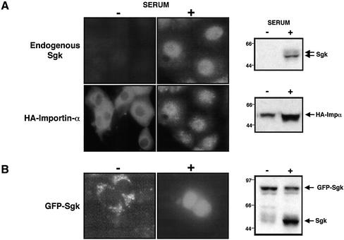
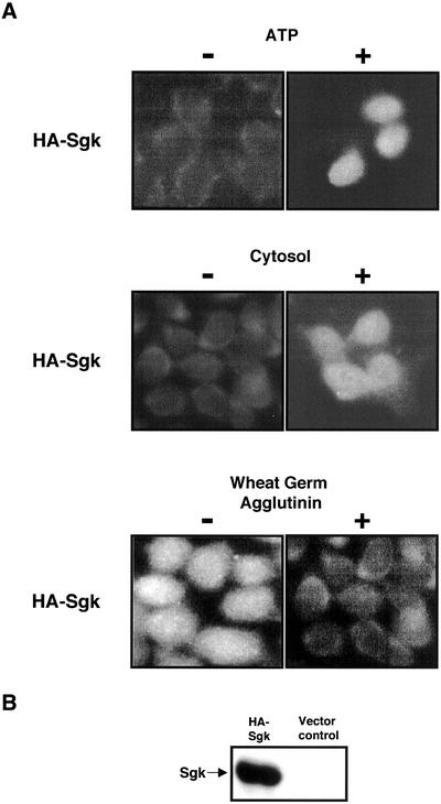
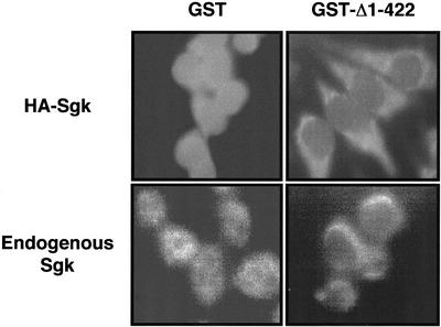
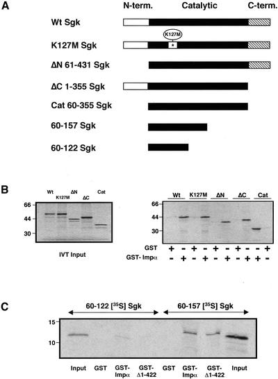
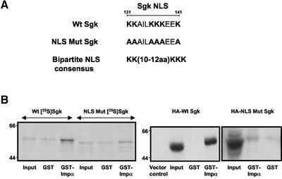
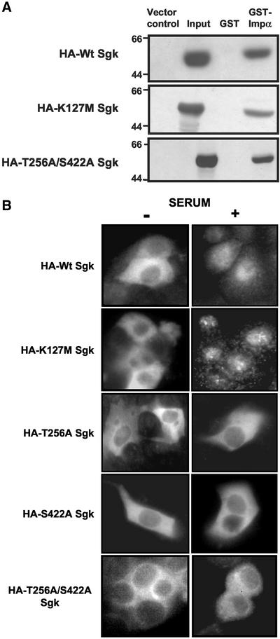
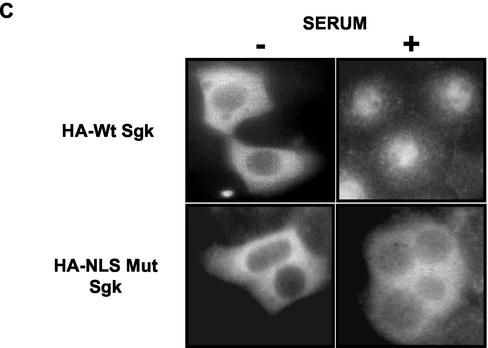
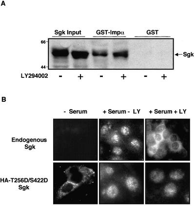
The transcriptionally regulated serum and glucocorticoid inducible protein kinase (Sgk) is localized to the nucleus in a serum-dependent manner, and a yeast two-hybrid genetic screen uncovered a specific interaction between Sgk and the importin-alpha nuclear import receptor. In vitro GST pull down assays demonstrated a strong and direct association of importin-alpha with endogenous Sgk and exogenously expressed HA-tagged Sgk, whereas both components coimmunoprecipitate and colocalize to the nucleus after serum stimulation. Consistent with an active mechanism of nuclear localization, the nuclear import of HA-Sgk in permeabilized cells required ATP, cytoplasm, and a functional nuclear pore complex. Ectopic addition of a 107 amino acid carboxy-terminal fragment of importin-alpha, which contains the Sgk binding region, competitively inhibited the ability of endogenous importin-alpha to import Sgk into nuclei in vitro. Mutagenesis of lysines by alanine substitution defined a KKAILKKKEEK sequence within the central domain of Sgk between amino acids 131-141 that functions as a nuclear localization signal (NLS) required for the in vitro interaction with importin-alpha and for nuclear import of full-length Sgk in cultured cells. The serum-induced nuclear import of Sgk requires the NLS-dependent recognition of Sgk by importin-alpha as well as the PI3-kinase-dependent phosphorylation of Sgk. Our results define a new role importin-alpha in the stimulus-dependent control of signal transduction by nuclear localized protein kinases.


Figures

Figure 1
Figure 1
Identification of importin-α-1 as a Sgk-interacting protein by yeast two-hybrid assay. (A) The yeast host strain EGY48 containing the Leu+ and LacZ reporter plasmids were transformed with the library derived importin-α clones 1, 2, and 3 along with one of three bait plasmids, pLexA-Sgk bait (top), pLexA-Lamin (middle), or pLexA-Bicoid (bottom). Individual Ura+ His+ Trp+ transformants were streaked onto synthetic plates containing galactose-raffinose based drop out media, lacking uracil, histidine, tryptophan, and leucine. The plates were incubated at 30°C for 3 d and the ability of individual bait proteins to interact with the library derived importin-α was determined by the growth of yeast on the selective medium. Similar results were obtained in two separate experiments. (B) The structure diagram of 529 amino acid full-length mouse importin-α (impα) depicts the N-terminal importin-β binding domain, 8–9 arm repeats traversing the central domain, and the carboxy -terminal enriched in acidic amino acids. The alignment of the truncated importin-α (Δ1–422) clone 1 isolated from the yeast two-hybrid screen is shown in comparison to the full-length importin-α (impα). The library derived importin-α is an N-terminal truncation that begins at amino acid 423 and continues to the C terminus at amino acid 529. Each of the three library derived clones displayed identical sequence homology and the size of encoded truncated protein only differed by 3–4 amino acids at the N-terminal truncation.
Figure 2
Figure 2
Analysis of Sgk and importin-α interactions by in vitro GST pull down assays. (A) Full-length Sgk cDNA was transcribed and translated in vitro with a rabbit reticulocyte lysate in the presence of [35S]methionine to produce [35S]Sgk. The SDS-PAGE and autoradiographic analysis of the unprogrammed lysate (UL) devoid of any Sgk cDNA is shown in the far left lane, and the programmed lysate representing an aliquot of the translated protein used for the binding assays is shown in the input lane. The in vitro–translated [35S]Sgk was incubated with glutathione sepharose beads bound with GST alone (GST), a fusion protein linked to the truncated importin-α (GST-Δ1–422), or with a GST-fusion protein linked to the full-length importin-α (GST-impα). Proteins retained on the beads were resolved by SDS-PAGE and visualized by autoradiography. The electrophoretic migration of the molecular weight standards in kDa are also shown. (B) GST pull down assays using the GST full-length importin-α fusion protein (GST-impα) were carried out with in vitro–translated [35S]Sgk, [35S]Jnk, or [35S]PKCζ and the proteins retained on the glutathione sepharose beads analyzed by SDS-PAGE and autoradiography. The in vitro–translated protein used in the assay is shown in the input lanes. (C) Subconfluent cultures of Con8.hd6 mammary epithelial tumor cells were serum starved for 72 h and then pulsed with 10% calf serum containing DME/F12 media for 4 h (+S), whereas control cultures were continued on serum-free media alone (−S). Cell extracts from serum-stimulated (+S) were incubated with either GST alone, with the full-length importin-α fusion protein GST-impα (left panel) or the truncated importin-α fusion protein GST-Δ1–422 (right panel) bound to glutathione sepharose beads. The bound proteins were separated on SDS-PAGE and immunoblotted with anti-Sgk antibodies to detect binding of the endogenous Sgk. As indicated in each panel, the input lanes show 10% of the cell lysate containing the endogenous Sgk included in the assay. Extracts from uninduced samples (−) are shown in the left panel. Arrows indicate migration of Sgk protein and the electrophoretic migration of molecular weight marker proteins are shown on the left side of each panel. Similar results were obtained in multiple experiments.
Figure 3
Figure 3
Coimmunoprecipitation of Sgk and importin-α. (A) Con8.hd6 mammary epithelial tumor cells were transfected with HA-impα expression vector (full-length importin-α) using lipofectamine method, and after serum starvation for 36 h, cells were stimulated for 4 h with 10% calf serum containing media to induce the production of endogenous Sgk. Precleared cell lysates were prepared as described in MATERIALS AND METHODS and immunoprecipitated with anti-Sgk antibodies (+) or with control rabbit IgG antibodies (−), and protein bound to the protein-A beads was fractionated by SDS-PAGE and immunoblotted with anti-HA monoclonal antibodies (right panel). Inputs (10%) reveal expression of HA-impα in the transfected samples compared with vector control lysates. The efficiency of Sgk immunoprecipitations were determined by immunoblotting the precleared lysates before (Pre) and after (Post) the Sgk-immunoprecipitation with anti-Sgk antibodies (left panel). (B) Hek 293 cells were transfected with expression plasmids encoding full-length importin-α (HA-impα) and the catalytic domain of Sgk (Cat Sgk), or with the empty vector controls. The cell lysates were prepared as detailed in MATERIALS AND METHODS and immunoprecipitated with anti-HA monoclonal antibodies (+) or with control mouse IgG antibodies (−), and Sgk bound to the protein-G beads was detected with anti-Sgk immunoblotting (right panel). Inputs (HA-impα and Cat Sgk) contain 10% of total protein and are compared with lysates prepared from vector transfected controls as indicated in each panel. Anti-HA immunoblotting of supernatants before (Pre) and after (Post) immunoprecipitating with anti-HA antibodies, to determine efficiency of immunoprecipitation of the HA-impα is shown in the left panel. The electrophoretic migration of molecular weight marker proteins is shown on the left side of each panel, and arrows indicate migration of HA-impα and heavy chain. Similar results were obtained from three independent experiments.
Figure 4
Figure 4
Subcellular localization of Sgk and importin-α. (A) Low confluent monolayers of Con8.hd6 mammary epithelial tumor cells grown on two-well Lab-Tek slides were transfected with expression vectors encoding the full-length HA-tagged importin-α (HA-impα) using lipofectamine. Cells were serum starved for 36 h, and then serum was boosted with 10% calf serum (+S) for 15 h. One set of cells was maintained under serum-free conditions for the duration of the experiment (−S). The colocalization of Sgk and transfected HA-importin-α was examined by double indirect immunofluorescence microscopy using anti-Sgk polyclonal antibodies and anti-HA monoclonal (1:1000, dilution) antibodies. The secondary antibodies used to detect the Sgk immune reaction was fluorescein isothiocyanate-conjugated goat anti-rabbit antibodies, whereas Texas-red/goat anti-mouse antibodies were used to detect the HA-importin-α. Protein expression of endogenous Sgk and exogenously expressed HA-importin-α in the treatment conditions used for colocalization studies were evaluated by immunoblotting with anti-Sgk or anti-HA antibodies, respectively. The electrophoretic migration of molecular weight marker proteins are shown on the left side of each gel and arrows indicate migration of Sgk and HA-impα. (B) Low confluent monolayers of Con8.hd6 mammary epithelial tumor cells grown on two-well Lab-Tek slides were transfected with an expression vector encoding the green fluorescence protein–tagged Sgk (GFP-Sgk) fusion protein. Cells were serum starved for 36 h, and then serum was boosted with 10% calf serum (+S) for 15 h. One set of cells was maintained under serum-free conditions for the duration of the experiment (−S). The localization of GFP-Sgk was monitored by direct fluorescence microscopy of formaldehyde-fixed cells using the FITC filter as detailed in MATERIALS AND METHODS. Protein expression of endogenous Sgk and exogenously expressed GFP-Sgk in the treatment conditions used for colocalization studies were evaluated by immunoblotting with anti-Sgk.
Figure 5
Figure 5
(A) Characteristics of the in vitro nuclear import of HA-Sgk. Hek 293 cells were transfected with expression vectors encoding the HA-Sgk, and S-100 cell cytosols prepared as described in MATERIALS AND METHODS. Nuclear import of the exogenously expressed Sgk proteins was examined in vitro either in the absence (−ATP) or presence of (+ATP) regenerating system (top), or in the absence (−) or presence (+) of cytosolic extract (middle), or with cytosol in the absence (−) or presence (+) of 0.5 mg/ml wheat germ agglutinin (bottom) using nuclei derived from permeabilized Hela cells. The import of HA-Sgk was assessed by indirect immunofluorescence microscopy using anti-HA antibodies as detailed in the text. (B) Expression of exogenously derived HA-Sgk in S-100 cytosols from Hek 293 cells as determined by anti-HA immunoblotting.
Figure 6
Figure 6
Carboxyterminal fragment of importin-α acts as a dominant negative molecule that inhibits in vitro Sgk nuclear import. Nuclear import reactions comprised of an intact ATP-regenerating system and cytosols containing either ectopically expressed HA-Sgk or serum-activated endogenous Sgk were carried out in the presence of purified GST alone or GST-Δ1–422 (4 μg each), and entry of the import substrates into permeabilized Hela nuclei was assessed by indirect immunofluorescence using anti-HA antibodies for HA-Sgk or polyclonal Sgk antibodies for endogenous Sgk.
Figure 7
Figure 7
Interaction of in vitro–translated Sgk mutants with recombinant full-length importin-α. (A) The structural diagrams are shown for the wild-type Sgk (Wt Sgk), the kinase dead mutant Sgk (K127 M) Sgk, an N-terminal deleted Sgk (ΔN 61–431 Sgk), a C-terminal deleted Sgk (ΔC 1–355 Sgk), a double truncation representing the central catalytic domain only of Sgk (Cat 60–355 Sgk), or two fragments of the central domain of Sgk (60–157 Sgk, 60–122 Sgk). (B) The [35S]methionine-labeled forms of the wild-type and indicated mutant Sgk proteins were synthesized using an in vitro translation system as described in Figure 2, and the resulting proteins (IVT inputs, left panel) were analyzed by SDS-PAGE and autoradiography. The in vitro–translated Sgk proteins (right panel) were incubated with either the GST protein alone or the GST full-length importin-α fusion protein (GST-impα) bound to glutathione sepharose beads, and the bound proteins were separated on SDS-PAGE and visualized by autoradiography. (C) GST pull down assays using the indicated fragments of the Sgk central catalytic domain were performed as above, except with the inclusion of truncated importin-α (GST-Δ1–422), in addition to the full-length importin-α (GST-impα) and GST alone. The electrophoretic migration of molecular weight markers are shown on the left side of each panel. Similar results were observed in three independent experiments.
Figure 8
Figure 8
Identification of the Sgk NLS required for binding to importin-α and serum-induced nuclear import. (A) Site-directed mutagenesis of the Sgk NLS. The Sgk NLS mutant was generated by substitution of the six highlighted lysines with alanines within the context of the full-length Sgk and is shown in comparison with the consensus bipartite NLS. (B) In vitro–translated wild-type Sgk (Wt [35S]Sgk) and NLS mutant Sgk (NLS Mut [35S]Sgk) proteins were incubated with either the GST protein alone or the GST-full-length importin-α fusion protein (GST-impα) bound to glutathione sepharose beads, and the bound proteins were separated on SDS-PAGE and visualized by autoradiography (left panel). Cell lysates prepared from Hek 293 cells transiently transfected with expression plasmids encoding HA-tagged forms of wild-type Sgk (HA-Wt Sgk) or NLS mutant Sgk (HA-NLS Mut Sgk) were incubated with GST protein alone or with the full-length importin-α fusion protein GST-IMPA bound to glutathione sepharose beads, and the proteins were retained on the beads fractionated by SDS-PAGE and immunoblotted with anti-HA antibodies (right panel). Inputs denote 10% of the total protein included in the binding reaction, and the electrophoretic migration of molecular weight marker proteins are shown on the left side of each panel. (C) Low confluent monolayers of Con8.hd6 mammary epithelial tumor cells grown on two-well Lab-Tek slides were transfected with expression vectors encoding either the wild-type HA-tagged Sgk (HA-Wt Sgk) or the NLS mutant Sgk (HA-NLS Mut Sgk) as in Figure 4. After serum starvation for 36 h, cells were serum boosted with 10% calf serum (+S) for 15 h, whereas the control slides remained on serum-free media (−S). The subcellular distribution of the wild-type and NLS mutant Sgk proteins were evaluated by indirect immunofluorescence microscopy using anti-HA monoclonal (1:1000, dilution) antibodies. Texas-red/goat anti-mouse antibodies was used as the secondary antibodies as in Figure 4 for detection of the HA-tagged proteins.
Figure 9
Figure 9
Characterization of Sgk phosphorylation and kinase mutants in the in vitro interaction with importin-α and in serum-induced nuclear translocation. (A) Hek 293 cells were transiently transfected with expression plasmids encoding HA-tagged forms of wild-type Sgk (HA-Wt Sgk), kinase dead Sgk (HA-K127 M Sgk), double phosphorylation site mutant of Sgk (HA- Sgk T256A/S422A), or with an empty expression vector (vector control) as described in the text. Cell lysates were incubated with GST protein alone or with the full-length importin-α fusion protein GST-impα bound to glutathione sepharose beads, and the proteins retained on the beads fractionated by SDS-PAGE and immunoblotted with anti-HA antibodies (left panels) as described in Figure 8. Inputs denote 10% of the total protein included in the binding reaction and are compared with vector-transfected controls. (B) Low confluent monolayers of Con8.hd6 mammary epithelial tumor cells were transfected with wild-type Sgk (HA-Wt Sgk), kinase dead Sgk (HA-K127 M Sgk), or with single phosphorylation site mutants (HA-T256A Sgk, HA-S422A Sgk) or a double phosphorylation site mutant (HA-T256A/S422A SGK) of Sgk. All constructs contained a HA epitope tag. Cells were serum starved for 36 h and then serum boosted with either 10% calf serum (+S) for 15 h or were continued on serum-free media (−S). The subcellular distribution of the Sgk proteins were assessed by indirect immunofluorescence microscopy using anti-HA monoclonal antibodies (right panels) as described in Figure 8.
Figure 9
Figure 9
Characterization of Sgk phosphorylation and kinase mutants in the in vitro interaction with importin-α and in serum-induced nuclear translocation. (A) Hek 293 cells were transiently transfected with expression plasmids encoding HA-tagged forms of wild-type Sgk (HA-Wt Sgk), kinase dead Sgk (HA-K127 M Sgk), double phosphorylation site mutant of Sgk (HA- Sgk T256A/S422A), or with an empty expression vector (vector control) as described in the text. Cell lysates were incubated with GST protein alone or with the full-length importin-α fusion protein GST-impα bound to glutathione sepharose beads, and the proteins retained on the beads fractionated by SDS-PAGE and immunoblotted with anti-HA antibodies (left panels) as described in Figure 8. Inputs denote 10% of the total protein included in the binding reaction and are compared with vector-transfected controls. (B) Low confluent monolayers of Con8.hd6 mammary epithelial tumor cells were transfected with wild-type Sgk (HA-Wt Sgk), kinase dead Sgk (HA-K127 M Sgk), or with single phosphorylation site mutants (HA-T256A Sgk, HA-S422A Sgk) or a double phosphorylation site mutant (HA-T256A/S422A SGK) of Sgk. All constructs contained a HA epitope tag. Cells were serum starved for 36 h and then serum boosted with either 10% calf serum (+S) for 15 h or were continued on serum-free media (−S). The subcellular distribution of the Sgk proteins were assessed by indirect immunofluorescence microscopy using anti-HA monoclonal antibodies (right panels) as described in Figure 8.
Figure 10
Figure 10
Effects of altering the PI 3-kinase–dependent phosphorylation of Sgk on the in vitro interaction with importin-α and in serum-induced nuclear translocation. (A) Subconfluent cultures of Con8.hd6 mammary epithelial cells were serum starved for 48 h and pretreated with or without 50 μM LY294002, a PI3-Kinase inhibitor, for 12 h. Cells were stimulated for 4 h with 10% calf-serum containing DMEM/F12 media either in the absence or presence of LY294002. Cell extracts from LY294002-treated (+) or untreated (−) cells were prepared as described in MATERIALS AND METHODS and incubated with GST protein alone or with the full-length importin-α fusion protein GST-Impα bound to glutathione sepharose beads, and the proteins were retained on the beads fractionated by SDS-PAGE and immunoblotted with anti-Sgk antibodies. Input lanes depict 10% of the extract prepared from LY294002-treated or untreated samples used in the binding assay (upper panel). (B) The nuclear localization of endogenous Sgk or exogenously expressed mutated Sgk with the T256 and S422 phosphorylation sites converted into aspartic acid (HA-T256D/S422D Sgk) and containing an HA epitope tag was assessed in Con8.hd6 mammary epithelial tumor cells. Low confluent monolayers of transfected and nontransfected cells were serum starved for 36 h, and then serum was boosted with 10% calf serum (+ Serum) for 15 h or maintained under the serum-free conditions (− Serum). Other sets of cells were pretreated with 50 μM LY294002 for 8 h and subsequently incubated with serum in the continued presence of LY294002 for another 15 h (+Serum +LY). The localization of endogenous Sgk was examined by indirect immunofluorescence microscopy using anti-Sgk polyclonal antibodies (lower panel) as described in Figure 4.

Similar articles

Cited by

References

    1. Adachi M, Fukuda M, Nishida E. Two co-existing mechanisms for nuclear import of MAP kinase: passive diffusion of a monomer and active transport of a dimer. EMBO J. 1999;18:5347–5358. - PMC - PubMed
    1. Adam SA, Marr RS, Gerace L. Nuclear protein import in permeabilized mammalian cells requires soluble cytoplasmic factors. J Cell Biol. 1990;111:807–816. - PMC - PubMed
    1. Alliston TN, Gonzalez-Robayna IJ, Buse P, Firestone GL, Richards JS. Expression and localization of serum/glucocorticoid-induced kinase in the rat ovary: relation to follicular growth and differentiation. Endocrinology. 2000;141:385–395. - PubMed
    1. Alliston TN, Maiyar AC, Buse P, Firestone GL, Richards JS. Follicle stimulating hormone-regulated expression of serum/glucocorticoid-inducible kinase in rat ovarian granulosa cells: a functional role for the Sp1 family in promoter activity. Mol Endocrinol. 1997;11:1934–1949. - PubMed
    1. Alvarez de la Rosa D, Zhang P, Naray-Fejes-Toth A, Fejes-Toth G, Canessa CM. The serum and glucocorticoid kinase sgk increases the abundance of epithelial sodium channels in the plasma membrane of Xenopus oocytes. J Biol Chem. 1999;274:37834–37839. - PubMed

Publication types

MeSH terms

LinkOut - more resources