. 2003 Apr;18(4):272-83.
doi: 10.1002/hbm.10084.

Functional somatotopy of finger representations in human primary motor cortex

Affiliations

Functional somatotopy of finger representations in human primary motor cortex

Peter Dechent et al. Hum Brain Mapp. 2003 Apr.

Abstract

To assess the degree of fine-scale somatotopy within the hand area of the human primary motor cortex (M1), functional mapping of individual movements of all fingers was performed in healthy young subjects (n = 7) using MRI at 0.8 x 0.8 mm2 resolution and 4 mm section thickness. The experimental design comprised both a direct paradigm contrasting single digit movements vs. motor rest and multiple differential paradigms contrasting single digit movements vs. the movement of another digit. Direct mapping resulted in largely overlapping activations. A somatotopic arrangement was only recognizable when considering the mean center-of-mass coordinates of individual digit representations averaged across subjects. In contrast, differential paradigms revealed more segregated and somatotopically ordered activations in single subjects. The use of center-of-mass coordinates yielded inter-digit distances ranging from 2.0 to 16.8 mm, which reached statistical significance for pairs of more distant digits. For the middle fingers, the functional somatotopy obtained by differential mapping was dependent on the choice of the digit used for control. These results confirm previous concepts that finger somatotopy in the human M1 hand area emerges as a functional predominance of individual digit representations sharing common areas in a distributed though ordered network.


Figures

Figure 1
Figure 1
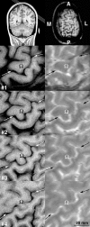
Top left: Coronal T1‐weighted MR image depicting the section orientation selected for functional mapping of the left‐hemispheric M1 hand area and (top right) corresponding oblique image (section 3, A = anterior, L = lateral, M = medial, P = posterior). (1–4: left) T1‐weighted FLASH images showing a magnified view of the M1 hand area (Ω) and (right) corresponding T2*‐weighted multi‐echo FLASH images used for functional mapping. The central sulcus is indicated by arrows.
Figure 2
Figure 2
Functional mapping of individual finger movements of the dominant right hand using a direct paradigm (i.e., D1 to D5 each vs. motor rest) in four consecutive sections covering the left‐hemispheric M1 (single subject). The overlap of activated areas within the M1 hand area reveals no clear somatotopy.
Figure 3
Figure 3
Functional mapping of individual finger movements of the dominant right hand using a differential paradigm (all possible combinations shown, e.g., D5 vs. D4, D5 vs. D3, D5 vs. D2, etc.) in a selected section (section 2, as in Fig. 2) of the left‐hemispheric M1 (same subject as in Fig. 2). The exact location of an individual finger representation strongly depends on the control digit used. Nevertheless, a somatotopic arrangement is recognizable.
Figure 4
Figure 4
Functional mapping of individual finger movements of the dominant right hand using a differential paradigm (selected combinations refer to D5 vs. D2, D4 vs. D1, D3 vs. D1, D2 vs. D3, and D1 vs. D5) in four consecutive sections covering the left‐hemispheric M1 (same subject as in Figs. 2 and 3). The segregation of individual finger representations reveals a somatotopic order.
Figure 5
Figure 5
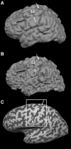
A: Surface reconstruction of the left hemisphere (same subject as in Figs. 2, 3, 4), which partially hides the M1 hand area (Ω) due to the convolution of the cerebral cortex. B: Segmented cortex at the level of the gray‐white matter boundary. C: Inflated visualization of the segmented cortex with former convex structures (“gyri”) shaded in light gray and former concave structures (“sulci”) shaded in dark gray. The white box corresponds to the region magnified in Figure 6; arrows indicate the central sulcus.
Figure 6
Figure 6
Individual finger representations in the left‐hemispheric M1 hand area on magnified views of the inflated cortex (same subject and same data as in Figs. 2 and 4, but without premotor and somatosensory activations). Top: Direct mapping of individual finger movements (i.e., D1 to D5 each vs. motor rest) results in activation areas with considerable overlap. Bottom: Differential mapping (selected combinations refer to D5 vs. D2, D4 vs. D1, D3 vs. D1, D2 vs. D3, and D1 vs. D5) yields more segregated and somatotopically arranged representations.
Figure 7
Figure 7
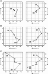
Somatotopic arrangement of individual finger representations in the left‐hemispheric M1 hand area viewed in a (left) posterior‐anterior and (right) inferior–superior projection (A = anterior, I = inferior, L = lateral, M = medial, P = posterior, S = superior). The coordinates (in mm) refer to mean center‐of‐mass activations (Table II) obtained for (A) a direct paradigm, (B) differential paradigms using all other digits, and (C) differential paradigms using directly neighboring digits only.
Figure 8
Figure 8
Reversible somatotopy of D3 and D4 finger representations in magnified views of the inflated cortex covering the left‐hemispheric M1 hand area (different subject as in Fig. 6). Top: Shifted activations obtained by differential mapping with neighboring fingers on either side (i.e., D3 vs. D2 and D3 vs. D5 as well as D4 vs. D3 and D4 vs. D5). Bottom: When referencing these activations against the representations of D2 (or D3), D3 (or D4) shows either a conventional (left) or reversed (right) somatotopic order.

Similar articles

Cited by

References

    1. Beisteiner R, Windischberger C, Lanzenberger R, Edward V, Cunnington R, Erdler M, Gartus A, Streibl B, Moser E, Deecke L (2001): Finger somatotopy in human motor cortex. Neuroimage 13: 1016–1026. - PubMed
    1. Dechent P, Frahm J (2000): Direct mapping of ocular dominance columns in human primary visual cortex. Neuroreport 11: 3247–3249. - PubMed
    1. Donoghue JP, Leibovic S, Sanes JN (1992): Organization of the forelimb area in squirrel monkey motor cortex: representation of digit, wrist, and elbow muscles. Exp Brain Res 89: 1–19. - PubMed
    1. Ferrier D. Experiments on the brain of monkeys. (1875): Proc R Soc London 23: 409–430.
    1. Foerster O (1936): The motor cortex of man in the light of Hughlings Jackson's doctrines. Brain 59: 135–159.

LinkOut - more resources