

Peptidergic innervation of human esophageal and cardiac carcinoma
- PMID: 12632484
- PMCID: PMC4621548
- DOI: 10.3748/wjg.v9.i3.399
Peptidergic innervation of human esophageal and cardiac carcinoma
Abstract
Aim: To investigate the distribution of neuropeptide-immunoreactive nerve fibers in esophageal and cardiac carcinoma as well as their relationship with tumor cells so as to explore if there is nerve innervation in esophageal and cardiac carcinoma.
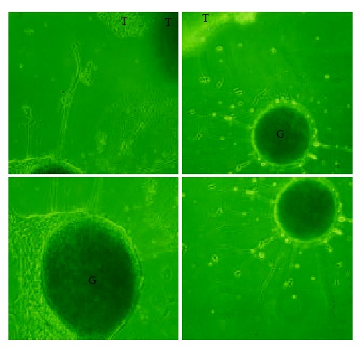
Methods: Esophageal and cardiac carcinoma specimens were collected from surgical operation. One part of them were fixed immediately with 4 % paraformaldehyde and then cut with a cryostat into 40-microm-thick sections to perform immunohistochemical analysis. Antibodies of ten kinds of neuropeptide including calcitonin gene-related peptide (CGRP), galanin (GAL), substance P (SP), etc. were used for immunostaining of nerve fibers. The other part of the tumor specimens were cut into little blocks (1 mm(3)) and co-cultured with chick embryo dorsal root ganglia (DRG) to investigate if the tumor blocks could induce the neurons of DRG to extend processes, so as to probe into the possible reasons for the nerve fibers growing into tumors.
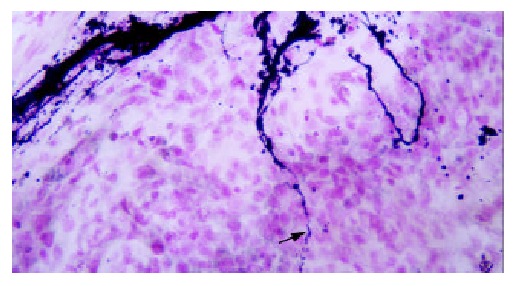
Results: Substantial amounts of neuropeptide including GAL-, NPY-, SP-immunoreactive nerve bundles and scattered nerve fibers were distributed in esophageal and cardiac carcinomas. The scattered nerve fibers waved their way among tumor cells and contacted with tumor cells closely. Some of them even encircled tumor cells. There were many varicosities aligned on the nerve fibers like beads. They were also closely related to tumor cells. In the co-culture group, about 63 % and 67 % of DRG co-cultured with esophageal and cardiac tumor blocks respectively extended enormous processes, especially on the side adjacent to the tumor, whereas in the control group (without tumor blocks), no processes grew out.
Conclusion: Esophageal and cardiac carcinomas may be innervated by peptidergic nerve fibers, and they can induce neurons of DRG to extend processes in vitro.
Figures


References
-
- Willis RA. The spread of the tumors in the human. 3rd ed. London: Butterworths. 1973:121–125.
-
- Luts L, Bergenfelz A, Alumets J, Sundler F. Peptide-containing nerve fibres in normal human parathyroid glands and in human parathyroid adenomas. Eur J Endocrinol. 1995;133:543–551. - PubMed
-
- Stack PS. Lymphomatous involvement of peripheral nerves: clinical and pathologic features. South Med J. 1991;84:512–514. - PubMed
-
- Takubo K, Takai A, Yamashita K, Yoshimatsu N, Kitano M, Sasajima K, Fujita K. Light and electron microscopic studies of perineural invasion by esophageal carcinoma. J Natl Cancer Inst. 1985;74:987–993. - PubMed
-
- Iishi H, Tatsuta M, Baba M, Uehara H, Nakaizumi A. Protection by galanin against gastric carcinogenesis induced by N-methyl-N'-nitro-N-nitrosoguanidine in Wistar rats. Cancer Res. 1994;54:3167–3170. - PubMed
Publication types
MeSH terms
Substances
LinkOut - more resources
Full Text Sources
Medical
Research Materials
Miscellaneous
