

A mouse model of severe acute pancreatitis induced with caerulein and lipopolysaccharide
- PMID: 12632523
- PMCID: PMC4621587
- DOI: 10.3748/wjg.v9.i3.584
A mouse model of severe acute pancreatitis induced with caerulein and lipopolysaccharide
Abstract
Aim: To establish a non-traumatic, easy to induce and reproducible mouse model of severe acute pancreatitis (SAP) induced with caerulein and lipopolyasccharide (LPS).
Methods: Thirty-two healthy mature NIH female mice were selected and divided at random into four groups (each of 8 mice), i.e., the control group (NS group), the caerulein group (Cn group), the lipopolysaccharide group (LPS group), and the caerulein+LPS group (Cn+LPS group). Mice were injected intraperitoneally with caerulein only, or LPS only, and caerulein and LPS in combination. All the animals were then killed by neck dislocation three hours after the last intraperitoneal injection. The pancreas and exo-pancreatic organs were then carefully removed for microscopic examination. And the pancreatic acinus was further observed under transmission electron microscope (TEM). Pancreatic weight, serum amylase, serum nitric oxide (NO) concentration, superoxide dismutase (SOD) and malondialdehyde (MDA) concentration of the pancreas were assayed respectively.
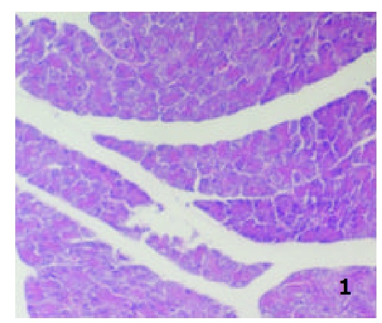
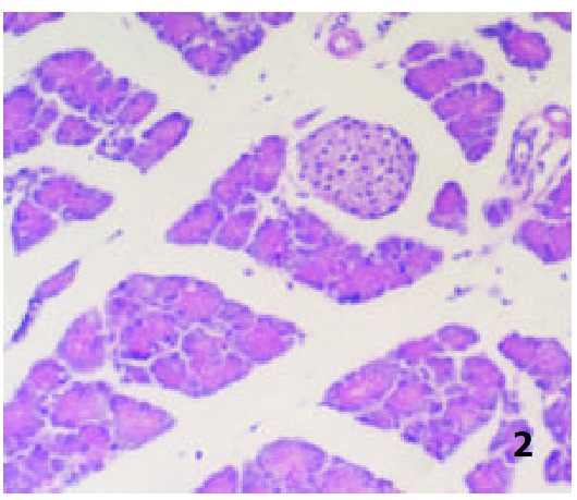
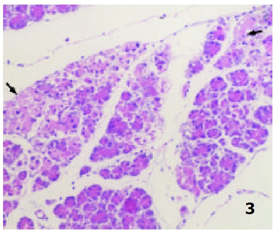
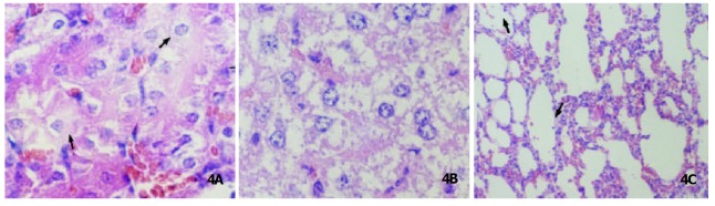
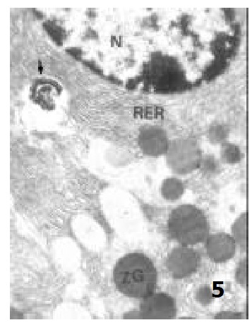
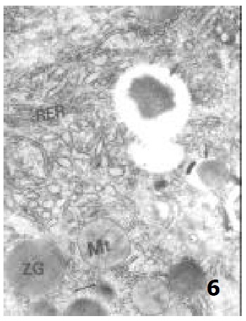
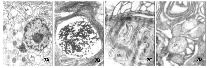
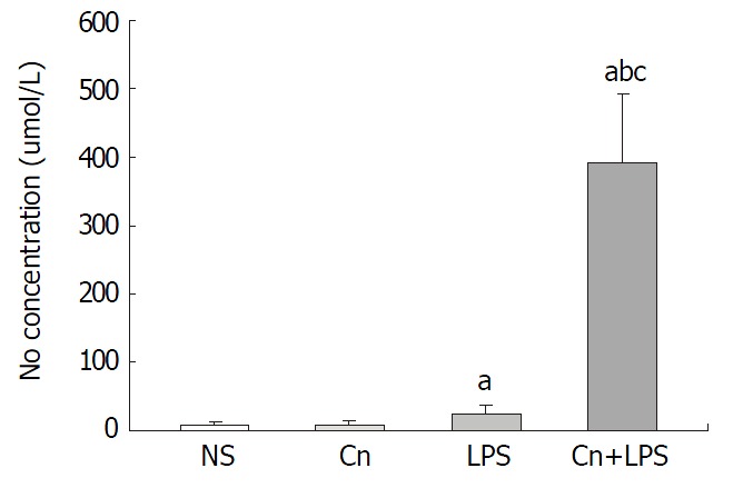
Results: (1) NS animals displayed normal pancreatic structure both in the exocrine and endocrine. In the LPS group, the pancreas was slightly edematous, with the infiltration of a few inflammatory cells and the necrosis of the adjacent fat tissues. All the animals of the Cn group showed distinct signs of a mild edematous pancreatitis characterized by interstitial edema, infiltration of neutrophil and mononuclear cells, but without obvious parenchyma necrosis and hemorrhage. In contrast, the Cn+LPS group showed more diffuse focal areas of nonviable pancreatic and hemorrhage as well as systemic organ dysfunction. According to Schmidt's criteria, the pancreatic histologic score showed that there existed significant difference in the Cn+LPS group in the interstitial edema, inflammatory infiltration, parenchyma necrosis and parenchyma homorrhage in comparison with those of the Cn group, LPS group and NS group (P<0.01 or P<0.05). (2) The ultrasturcture of acinar cells was seriously damaged in the Cn+LPS group. Chromatin margination of nuclei was present, the number and volume of vacuoles greatly increased. Zymogen granules (ZGs) were greatly decreased in number and endoplasmic reticulum exhibited whorls. The swollen mitochondria appeared, the crista of which was decreased in number or disappeared. (3) Pancreatic weight and serum amylase levels in the Cn +LPS was significantly higher than those of the NS group and the LPS group respectively (P<0.01 or P<0.05). However, the pancreatic wet weight and serum amylase concentration showed no significant difference between the Cn+LPS group and the Cn group. (4) NO concentration in the Cn+LPS group was significantly higher than that of NS group, LPS group and Cn group(P<0.05 or P<0.01). (5) The SOD and MDA concentration of the pancreas in the Cn+LPS group were significantly higher than those of NS, LPS and Cn groups (P<0.05 or P<0.01).
Conclusion: The mouse model of severe acute pancreatitis could be induced with caerulein and LPS, which could be non-traumatic and easy to induce, reproducible with the same pathological characteristics as those of SAP in human, and could be used in the research on the mechanism of human SAP.
Figures
References
-
- Baron TH, Morgan DE. Acute necrotizing pancreatitis. N Engl J Med. 1999;340:1412–1417. - PubMed
-
- Pastor CM, Frossard JL. Are genetically modified mice useful for the understanding of acute pancreatitis. FASEB J. 2001;15:893–897. - PubMed
-
- Lecesne R, Taourel P, Bret PM, Atri M, Reinhold C. Acute pancreatitis: interobserver agreement and correlation of CT and MR cholangiopancreatography with outcome. Radiology. 1999;211:727–735. - PubMed
MeSH terms
Substances
LinkOut - more resources
Full Text Sources
Medical
Miscellaneous
