. 2003 Apr;62(4):297-302.
doi: 10.1136/ard.62.4.297.

Anti-inflammatory effects of leflunomide on cultured synovial macrophages from patients with rheumatoid arthritis

Affiliations

Anti-inflammatory effects of leflunomide on cultured synovial macrophages from patients with rheumatoid arthritis

M Cutolo et al. Ann Rheum Dis. 2003 Apr.

Abstract

Background: Leflunomide and its active metabolite A77 1726 reversibly inhibits the enzyme dihydro-orotate dehydrogenase, the rate limiting step in de novo synthesis of pyrimidines and progression of the cell cycle in different cell lines, mainly activated T lymphocytes.

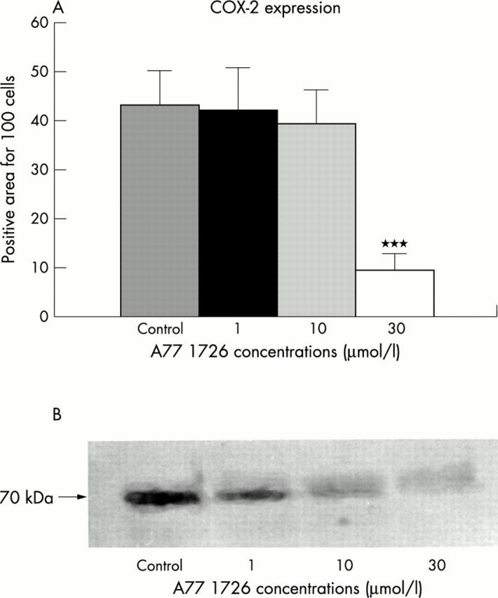
Objective: To analyse in vitro the possible anti-inflammatory effects exerted by A77 1726, on cultured macrophages, obtained from the synovial tissues of patients with rheumatoid arthritis (RA).

Methods: The effects of different doses of A77 1726 on intracytoplasmic expression and extracellular concentration of inflammatory cytokines (tumour necrosis factor alpha (TNFalpha), interleukin (IL) 1beta, IL6), as well as the influence on production and expression of intercellular adhesion molecule-1 (ICAM-1) and cyclo-oxygenase 2 (COX-2) by primary cultures of synovial macrophages from patients with RA, were evaluated by immunocytochemistry and western blot analysis. The observations were made at four and 24 hours.

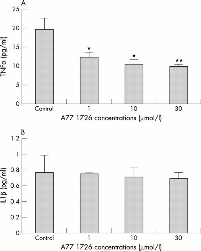
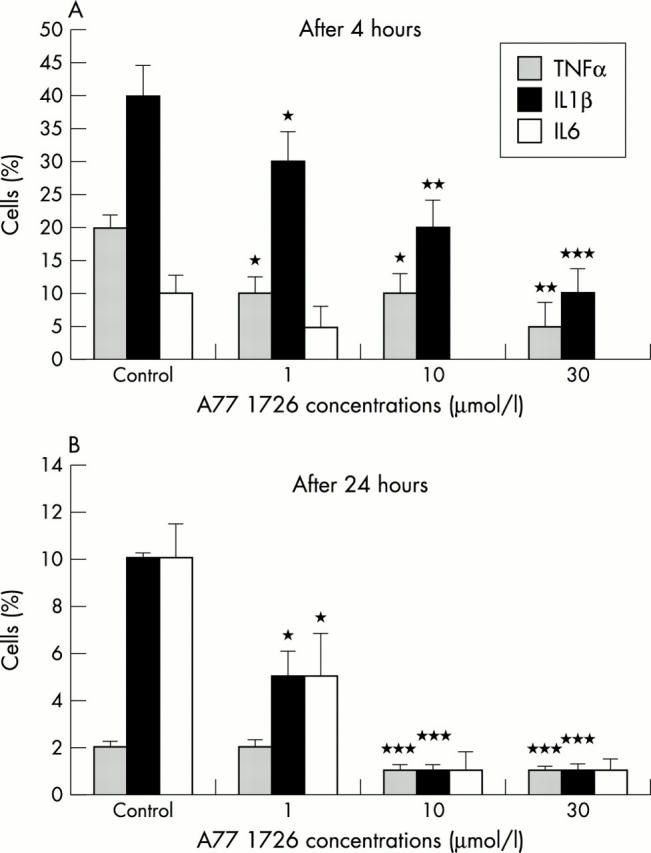
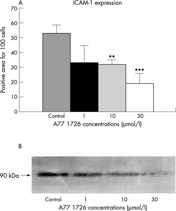
Results: A progressive and significant time and dose dependent decrease of the number of positive macrophages for intracellular TNFalpha and IL1beta, treated with different doses of A77 1726, was found in comparison with untreated cells. The extracellular concentration of TNFalpha was found to be significantly decreased in media containing cultured macrophages at 24 hours for all tested doses of A77 1726. At 24 hours, a significant time and dose dependent decrease of ICAM-1 and COX-2 expression by cultured macrophages after A77 1726 treatment was found.

Conclusions: In conclusion, the mechanism of antiproliferative activity exerted by leflunomide on activated T lymphocytes seems to be the same mechanism (alteration of the cell cycle progression) which interferes with the functions of other activated cells-namely, the monocytes/macrophages, which are strongly involved in the inflammatory reaction in RA synovial tissue. The positive clinical results seem to confirm that leflunomide exerts an anti-inflammatory action on phagocytic cells in short and long term treatment of RA.


Figures

Figure 1
Figure 1
(A) TNFα concentrations at 24 hours in culture media after incubation of RA synovial macrophages with A77 1726 at concentrations of 1, 10, and 30 µmol/l. *p<0.05; **p<0.01 versus controls. (B) IL1ß concentrations at 24 hours in culture media after incubation of RA synovial macrophages with A77 1726 at concentrations of 1, 10, and 30 µmol/l. No significant changes were found.
Figure 2
Figure 2
(A) Intracytoplasmic expression of TNFα, IL1ß and IL6 in RA synovial macrophages treated with different concentrations of A77 1726 after four hours. The percentage of TNFα and IL1ß positive cells decreased significantly in a dose dependent manner. *p<0.05; **p<0.01; ***p<0.001 versus controls. IL6 was poorly expressed and the effects of A77 1726 were not significant. (B) Intracytoplasmic expression of TNFα, IL1ß, and IL6 in RA synovial macrophage cells with different concentrations of A77 1726 after 24 hours. The percentage of IL1ß and IL6 positive cells decreased significantly in a dose dependent manner. *p<0.05; ***p<0.01 versus controls. TNFα was poorly expressed at 24 hours and the effect of A77 1726 was not significant.
Figure 5
Figure 5
COX-2 expression by cultured RA synovial macrophages with (A) and without (B) treatment with A77 1726 (30 µmol/l). ICAM-1 expression by cultured RA synovial macrophages with (C) and without (D) treatment with A77 1726 (30 µmol/l). Intracytoplasmic IL1 expression by cultured RA synovial macrophages with (E) and without (F) treatment with A77 1726 (30 µmol/l).
Figure 3
Figure 3
(A) ICAM-1 expression in RA synovial macrophages treated with different concentrations of A77 1726 after 24 hours as evaluated by immunocytochemistry and image analysis. The staining positivity was expressed as a percentage for cell number. **p<0.01; ***p<0.001. (B) ICAM-1 expression in RA synovial macrophages treated with different concentrations of A77 1726 after 24 hours as evaluated by western blot analysis.
Figure 4
Figure 4
(A) COX-2 expression in RA synovial macrophages treated with different concentrations of A77 1726 after 24 hours as evaluated by immunocytochemistry and image analysis. The staining positivity was expressed as a percentage for cell number. ***p<0.001. (B) COX-2 expression in RA synovial macrophages treated with different concentrations of A77 1726 after 24 hours as evaluated by western blot analysis.

References

    1. Ann Rheum Dis. 2001 Aug;60(8):729-35 - PubMed
    1. Rheumatology (Oxford). 2001 Apr;40(4):367-74 - PubMed
    1. Ann N Y Acad Sci. 2002 Jun;966:232-7 - PubMed
    1. Annu Rev Biochem. 1980;49:253-79 - PubMed
    1. Eur J Immunol. 1990 Jul;20(7):1591-6 - PubMed

MeSH terms