Microarchitecture and protective mechanisms in synovial tissue from clinically and arthroscopically normal knee joints
- PMID: 12634226
- PMCID: PMC1754505
- DOI: 10.1136/ard.62.4.303
Microarchitecture and protective mechanisms in synovial tissue from clinically and arthroscopically normal knee joints
Abstract
Background: Synovial biopsies are used to study synovial immunopathology and are increasingly applied for the evaluation of new therapeutic strategies in chronic arthritis. Therefore, it is essential to be informed on the complete spectrum of synovial immunopathology.
Objective: To describe the cellular content, cytokine and cell adhesion molecule expression in synovial tissue from clinically and arthroscopically normal knees.
Methods: Synovial tissue was obtained from 20 normal subjects at the time of knee joint arthroscopy for unexplained knee pain. Tissue sections were studied for basic histopathology and for a range of cell surface markers, cytokines, and cell adhesion molecules by immunoperoxidase staining. Stained sections were evaluated by semiquantitative scoring and digital image analysis.
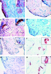
Results: Normal synovial tissue is composed predominantly of fibrofatty areolar tissue, with a variable thickness of intimal lining, composed of both CD68 positive macrophages and CD55 positive fibroblast-like synoviocytes. Interleukin 1 receptor antagonist (IL1Ra) was frequently detected in the synovial membrane of normal subjects (mean (SD) integrated optical density (IOD)=3809.6 (3893.9)), but both tumour necrosis factor alpha (TNFalpha) and interleukin 1beta (IL1beta) were rarely detected. In addition, cell adhesion molecules were rarely detected in the normal synovial membrane, with the exception of intercellular cell adhesion molecule-1 (ICAM-1) and vascular cell adhesion molecule-1 (VCAM-1). Osteoprotegerin (OPG) expression was abundant on synovial lining macrophages (mean (SD) IOD=5276 (4716) as well as endothelial cells (mean (SD) IOD=557 (226)), but receptor activator of nuclear factor kappa ligand (RANKL) expression was rarely seen.
Conclusions: The normal synovial membrane has a variable architecture, including thickness of the lining and the subintimal cell infiltrate, with little inflammatory cytokine production or expression of cell adhesion molecules. The excess of OPG expression over RANKL and IL1Ra over IL1 may be important for protection against joint damage
Figures

References
MeSH terms
Substances
LinkOut - more resources
Full Text Sources
Miscellaneous

