. 2003 Apr;77(7):3939-49.
doi: 10.1128/jvi.77.7.3939-3949.2003.

The leader of human immunodeficiency virus type 1 genomic RNA harbors an internal ribosome entry segment that is active during the G2/M phase of the cell cycle

Affiliations

The leader of human immunodeficiency virus type 1 genomic RNA harbors an internal ribosome entry segment that is active during the G2/M phase of the cell cycle

Ann Brasey et al. J Virol. 2003 Apr.

Abstract

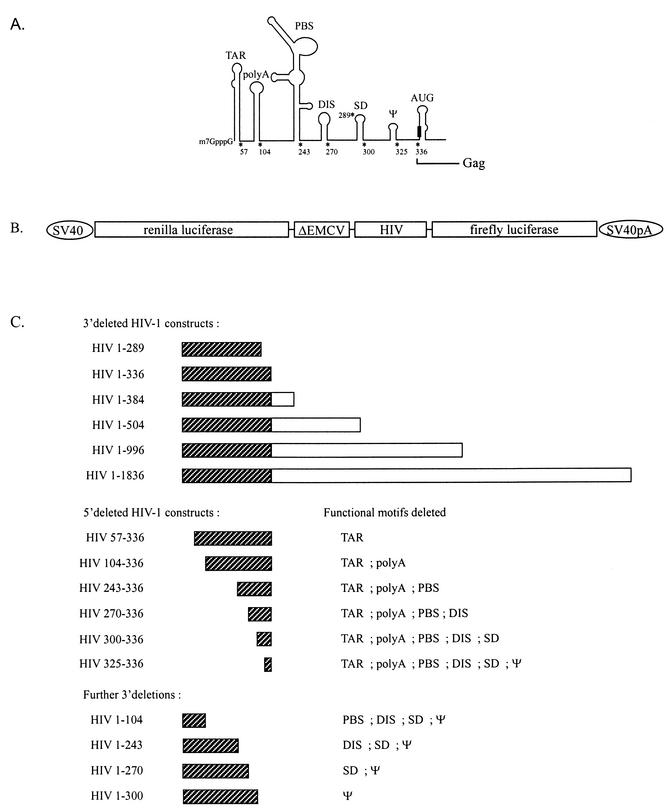
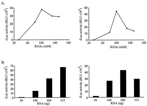
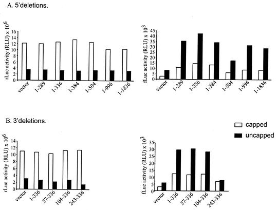
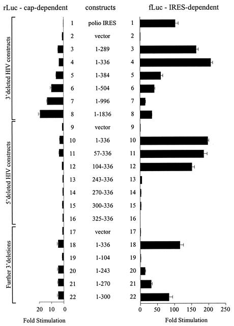
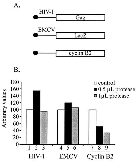
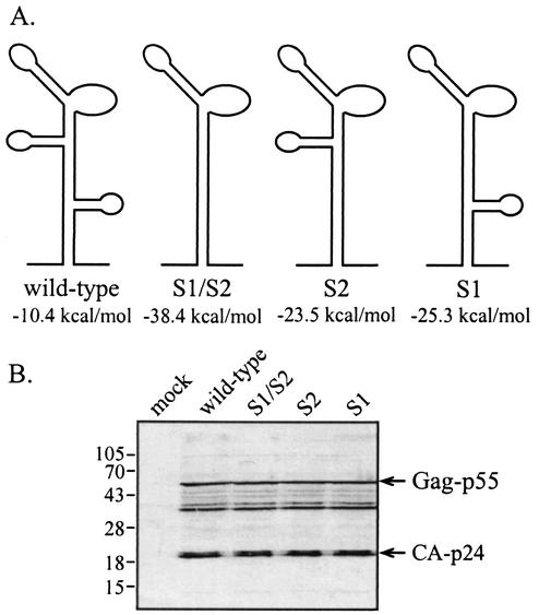
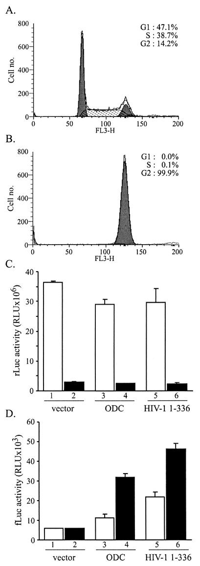
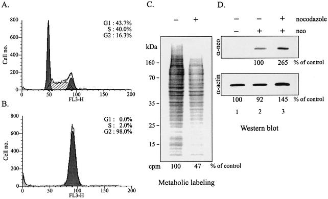
The 5' leader of the human immunodeficiency virus type 1 (HIV-1) genomic RNA contains highly structured domains involved in key steps of the viral life cycle. These RNA domains inhibit cap-dependent protein synthesis. Here we report that the HIV-1 5' leader harbors an internal ribosome entry site (IRES) capable of driving protein synthesis during the G(2)/M cell cycle phase in which cap-dependent initiation is inhibited. The HIV-1 IRES was delineated with bicistronic mRNAs in in vitro and ex vivo assays. The HIV-1 leader IRES spans nucleotides 104 to 336 and partially overlaps the major determinants of genomic RNA packaging. These data strongly suggest that, as for HIV-1 transcription, IRES-mediated translation initiation could play an important role in virus replication during virus-induced G(2)/M cell cycle arrest.


Figures

FIG. 1.
FIG. 1.
Design of bicistronic HIV-1 plasmids. (A) Schematic representation of the functional domains in the HIV-1 5′ leader. The RNA 5′ leader region contains the trans-activation-responsive region (TAR), the poly(A) hairpin, the primer binding site (PBS), the dimer initiation site (DIS), the major splice donor (SD), the core packaging signal (ψ), and a hairpin containing the initiator AUG (AUG loop). The asterisks denote the start and end points of deletion mutations that were used in this study. The black box represents the initiator AUG. Adapted from Berkhout (10). (B) Schematic representation of the dual luciferase reporter plasmid used in this study. The different HIV-1 fragments were subcloned into the intercistronic region as described in Materials and Methods. ΔEMCV is the encephalomyocarditis virus IRES which contains an inhibitory deletion (24, 83). (C) PCR-generated fragments of HIV-1 used in this study. The hatched boxes represent leader sequences; the white boxes represent gag coding region sequences.
FIG. 2.
FIG. 2.
Optimization of a HeLa cell in vitro translation system. Effect of potassium acetate (KOAc, A) on translation of Renilla (rLuc) and firefly luciferase (fLuc) ORFs (first and second cistrons, respectively). RLU, relative light units. (B) RNA dose-response. The RNA contains the full-length HIV-1 5′ leader (nucleotides 1 to 336).
FIG. 3.
FIG. 3.
HIV-1 IRES activity in a HeLa in vitro translation extract. Bicistronic capped and uncapped mRNAs were translated in a HeLa extract as described in Materials and Methods. Renilla luciferase and firefly luciferase activities (left and right panels, respectively) of the 3′-deleted HIV-1 constructs (A) and part of the 5′-deleted constructs (B) are shown. The results are from a representative experiment. See the legend to Fig. 2 for abbreviations.
FIG. 4.
FIG. 4.
Demarcation of the HIV-1 IRES. Renilla luciferase (rLuc) and firefly luciferase (fLuc) activity (left and right panels, respectively) in HeLa cells 28 h after transient cotransfection with the LacZ reporter and HIV-1 constructs. The results are from a representative experiment performed in triplicate and are expressed as stimulation of translation relative to the control vector. The results did not vary by more than 10%.
FIG. 5.
FIG. 5.
HIV-1 IRES-driven translation functions with proteolytically cleaved eIF4G. Monocistronic capped RNAs from HIV-1, encephalomyocarditis virus (EMCV), and cyclin B2 (A) were in vitro translated in a rabbit reticulocyte lysate treated with the foot-and-mouth disease virus L protease as previously described (52, 53, 62, 64). [35S]methionine-labeled proteins were subjected to SDS-PAGE and quantified (B) as described in Materials and Methods.
FIG. 6.
FIG. 6.
Increased secondary structure in the primer-binding site region does not inhibit translation. C33A cells were transfected with the wild-type or mutant proviral constructs shown in panel A. Total cellular extracts were prepared 3 days posttransfection, and viral proteins were detected by Western blotting with serum from an HIV-1-infected individual (B). The positions of the HIV-1 gag-p55 precursor protein and the mature CA-p24 protein are indicated on the right. Positions of molecular size markers are indicated on the left (in kilodaltons).
FIG. 7.
FIG. 7.
HIV-1 IRES is active in translation extracts prepared from mitotic cells. FACS analysis of untreated (A) and nocodazole-treated (B) HeLa cells from which in vitro translation extracts were prepared.In vitro translation of bicistronic capped mRNA in untreated (□) and nocodazole-treated (▪) HeLa cell extracts (C and D). The values are averages from two independent experiments. The difference in the translation efficiencies of the extracts was normalized by setting the firefly luciferase activity of the negative control vector plasmid to an equal value. See the legend to Fig. 2 for abbreviations.
FIG. 8.
FIG. 8.
HIV-1 IRES functions ex vivo in G2/M-arrested HeLa cells. FACS analysis of untreated (A) and nocodazole-treated (B) HeLa cells which express Plap/neo as described in Materials and Methods. (C) [35S]methionine-labeled proteins were resolved by SDS-PAGE and visualized by autoradiography. (D) Western blotting for neomycin and actin proteins was performed as described in Materials and Methods. Experiments were repeated three times, and the results of a representative experiment are shown. The results did not differ by more than 20%.

References

    1. Abbink, T. E., and B. Berkhout. A novel long-distance base-pairing interaction in human immunodeficiency virus type 1 RNA occludes the gag start codon. J. Biol. Chem., in press. - PubMed
    1. Adachi, A., H. E. Gendelman, S. Koenig, T. Folks, R. Willey, A. Rabson, and M. A. Martin. 1986. Production of acquired immunodeficiency syndrome-associated retrovirus in human and nonhuman cells transfected with an infectious molecular clone. J. Virol. 59:284-291. - PMC - PubMed
    1. Aldovini, A., and R. A. Young. 1990. Mutations of RNA and protein sequences involved in human immunodeficiency virus type 1 packaging result in production of noninfectious virus. J. Virol. 64:1920-1926. - PMC - PubMed
    1. Alizon, M., S. Wain-Hobson, L. Montagnier, and P. Sonigo. 1986. Genetic variability of the AIDS virus: nucleotide sequence analysis of two isolates from African patients. Cell 46:63-74. - PubMed
    1. Attal, J., M. C. Theron, F. Taboit, M. Cajero-Juarez, G. Kann, P. Bolifraud, and L. M. Houdebine. 1996. The RU5 ('R') region from human leukaemia viruses (HTLV-1) contains an internal ribosome entry site (IRES)-like sequence. FEBS Lett. 392:220-224. - PubMed

Publication types

LinkOut - more resources