. 2003 Apr;77(7):4033-42.
doi: 10.1128/jvi.77.7.4033-4042.2003.

Tumor necrosis factor alpha inhibition of hepatitis B virus replication involves disruption of capsid Integrity through activation of NF-kappaB

Affiliations

Tumor necrosis factor alpha inhibition of hepatitis B virus replication involves disruption of capsid Integrity through activation of NF-kappaB

Michael Biermer et al. J Virol. 2003 Apr.

Abstract

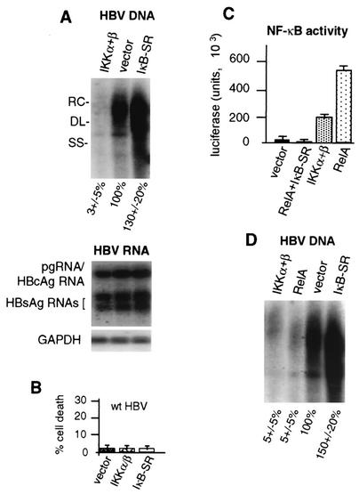
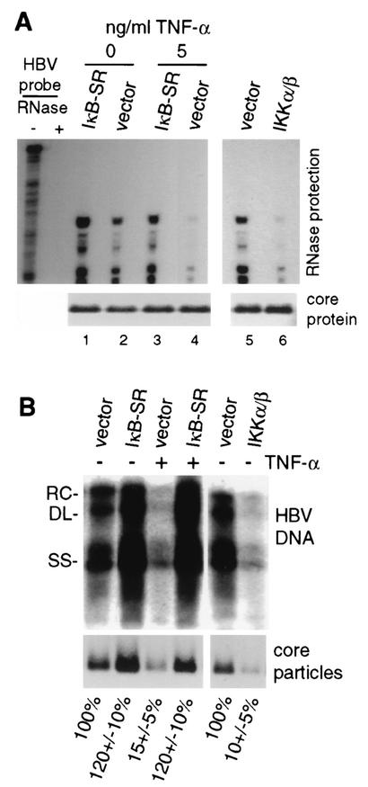
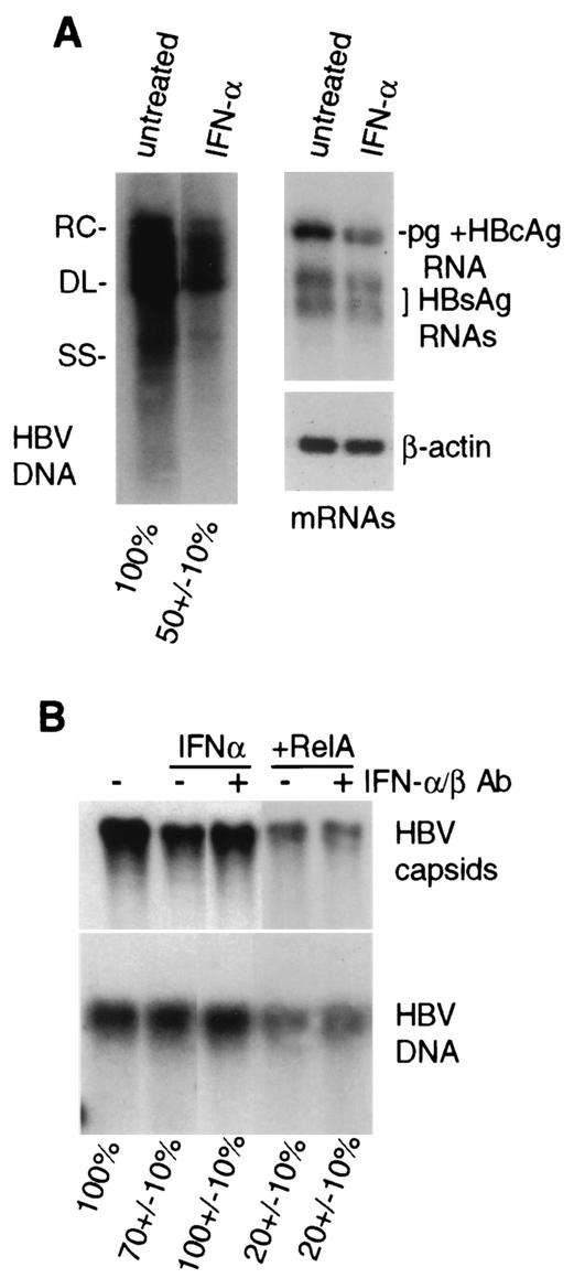
Chronic infection by hepatitis B virus results from an inability to clear the virus, which is associated with liver disease and liver cancer. Tumor necrosis factor alpha (TNF-alpha) is associated with noncytopathic clearance of hepatitis B virus in animal models. Here we demonstrate that the nuclear factor kappaB (NF-kappaB) signaling pathway is a central mediator of inhibition of hepatitis B virus by TNF-alpha and we describe the molecular mechanism. TNF-alpha is shown to suppress hepatitis B virus DNA replication without cell killing by disrupting the formation or stability of cytoplasmic viral capsids through a pathway requiring the NF-kappaB-activating inhibitor of kappaB kinase complex IKK-alpha/beta and active transcription factor NF-kappaB. Hepatitis B virus replication could also be inhibited and viral capsid formation could be disrupted in the absence of TNF-alpha solely by overexpression of IKK-alpha/beta or strong activation of NF-kappaB. In contrast, inhibition of NF-kappaB signaling stimulated viral replication, demonstrating that HBV replication is both positively and negatively regulated by the level of activity of the NF-kappaB pathway. Studies are presented that exclude the possibility that HBV inhibition by NF-kappaB is carried out by secondary production of gamma interferon or alpha/beta interferon. These results identify a novel mechanism for noncytopathic suppression of hepatitis B virus replication that is mediated by the NF-kappaB signaling pathway and activated by TNF-alpha.


Figures

FIG. 1.
FIG. 1.
HBV replication and transcription in HepG2 cells. (A) Schematic representation of replication-competent HBV genomic DNA. Shown is a head-to-tail replicon of 1.2 copies (1.2-mer) of the HBV genome. Indicated are the duplicated viral core protein (HBc) coding regions, the single HBx coding region, the polymerase (pol) coding region, and the base pair junction of HBV strain ayw. (B) Southern blot analysis of cytoplasmic HBV core particle-associated viral genomic DNA from equal numbers of cells transfected with wild-type HBV, the HBV HBx mutant, and the mutant trans complemented with an HBx expression vector for 4 days. Replicative DNA intermediates correspond to RC (relaxed circular), DL (double-stranded linear), and SS (single-stranded linear) DNAs. A Northern blot analysis of viral pregenomic (pg), core (HBc), and envelope (HBsAg) mRNAs and cellular GAPDH mRNA obtained from equal numbers of cells in duplicate plates is shown. Quantification of three independent experiments was obtained by densitometry and is shown at the bottom. Data were normalized to those obtained with the wild-type (wt) HBV sample.
FIG. 2.
FIG. 2.
Effect of TNF-α on HBV replication and cell viability. HepG2 cells were transfected with HBV 1.2-mer genomic DNA and treated with TNF-α for 4 days, with daily replenishment at the indicated doses. (A) Cytoplasmic core particles were purified from equal numbers of cells, and associated HBV DNA was extracted and detected by Southern blot analysis. Poly(A)+ mRNA was selected and detected by Northern blot analysis from duplicate plates of cells. RC, relaxed circular; DL, double stranded linear; SS, single stranded linear. (B) HepG2 cells were transfected with the vector alone or with wild-type HBV genomic DNA, with or without treatment with 5 ng of TNF-α per ml. At 4 days posttransfection, cells were assayed for percent death by the LDH assay. Untransfected cells were treated with 5 ng of TNF-α per ml and 10 μg of cycloheximide (Cyclo.) per ml for 10 h and then assayed for cell death. Results of both experiments were averaged from three independent assays and quantified by densitometry. Data were normalized to the untreated sample.
FIG. 3.
FIG. 3.
Role of NF-κB inhibition in TNF-α suppression of HBV replication. HepG2 cells were transfected with HBV 1.2-mer genomic DNA for 1 day and then treated for 4 days with the indicated dose of TNF-α. An expression plasmid for the IκB-SR superrepressor of NF-κB was included in the initial transfection as indicated. Cytoplasmic viral core particles were purified from equal numbers of cells, and associated HBV DNA was detected by Southern blot analysis. Northern blot analysis was done with poly(A)+ RNA extracted from equal numbers of cells in duplicate plates. Autoradiograms were quantified by densitometry of at least three independent experiments and normalized to the untreated control without IκB-SR (first lane). RC, relaxed circular; DL, double stranded linear; SS, single stranded linear.
FIG. 4.
FIG. 4.
Characterization of NF-κB activation by TNF-α or during HBV replication. HepG2 cells were either nontransfected (nontransf.) or transfected with wild-type or HBx mutant HBV genomic DNA for 3 days. Cells were treated with 5 ng of TNF-α per ml for 30 min as indicated. Nuclear extracts were prepared, and equal amounts (10 μg) were used to assay NF-κB DNA binding activity by EMSA with a 32P-labeled double-stranded oligonucleotide probe containing a single NF-κB binding site. Unlabeled-competitor (cold comp.) ablation of NF-κB binding was performed by using a 100-fold molar excess of unlabeled NF-κB oligonucleotide. Complexes were resolved by electrophoresis and autoradiography as described previously (56). Complexes were quantified by densitometry.
FIG. 5.
FIG. 5.
Role of NF-κB pathway in suppression of HBV replication. HepG2 cells were transfected with wild-type 1.2-mer HBV genomic DNA and the vector alone or the vector expressing NF-κB-activating kinase IKK-α/β or the NF-κB superrepressor IκB-SR. (A) Southern blot analysis on HBV core particle-associated viral DNA, and Northern blot analysis was performed on poly(A)+ RNA obtained from equal numbers of cells 4 days posttransfection in duplicate plates. RC, relaxed circular; DL, double stranded linear; SS, single stranded linear. (B) HepG2 cells were cotransfected with wild-type (wt) HBV genomic DNA and the vector expressing IKK-α/β or IκB-SR or the vector alone and analyzed for the level of cell death by the LDH assay at 3 days posttransfection. The data shown are averages of three independent experiments. (C) HepG2 cells were transfected with an NF-κB-dependent luciferase transcriptional reporter and the vector alone, the RelA NF-κB subunit expression vector, the vector expressing IKK-α/β, or the vectors expressing RelA and IκB-SR. NF-κB activity was determined by three independent luciferase assays using equal amounts of cellular extracts at 3 days posttransfection. (D) HepG2 cells were transfected with wild-type HBV genomic DNA and the expression vector for IKK-α/β or RelA, the vector alone, or IκB-SR. Viral core particles were isolated from equal numbers of cells at 4 days posttransfection, and viral DNA was assayed by Southern blot analysis. Autoradiograms were quantified by densitometry of at least three independent experiments and normalized to vector-plus-HBV samples.
FIG. 6.
FIG. 6.
Effects of TNF-α and NF-κB on HBV replication, core protein, and particles. (A) HepG2 cells were transfected with wild-type HBV genomic DNA and the vector alone or the expression vector for IKK-α/β or IκB-SR for 4 days. Cytoplasmic core particles were purified from equal numbers of cells, and RNase protection analysis was carried out on the 5′ end of the encapsidated pgRNA. Protected RNA fragments were resolved by denaturing urea-acrylamide gel electrophoresis and autoradiography. Total core protein was determined by SDS-polyacrylamide gel electrophoresis of equal amounts of cellular lysates and detected by immunoblot analysis with antibodies to HBcAg. TNF-α treatment was performed for 1 to 4 days with daily replenishment. (B) HepG2 cells were transfected and left untreated or treated with 5 ng of TNF-α per ml as described above. Southern blot analysis was conducted on HBV DNA extracted from core particles obtained from equal numbers of cells. Core particles were resolved by native agarose gel electrophoresis using the same cell lysates examined for HBV replication and subjected to immunoblot analysis with antibodies to HBcAg. See Materials and Methods for details. Results were quantified by densitometry of at least three independent autoradiograms and normalized to the untreated vector control. RC, relaxed circular; DL, double stranded linear; SS, single stranded linear.
FIG. 7.
FIG. 7.
IFNs are not the mediators of NF-κB inhibition of HBV replication. (A) HepG2 cells were transfected with HBV genomic DNA and 1 day later mock treated or treated with 1,000 U of recombinant IFN-α per ml for 3 days, with daily replenishment. Core-associated viral DNA and poly(A)+ mRNA Northern analyses were done with equal numbers of cells from duplicate plates. RC, relaxed circular; DL, double stranded linear; SS, single stranded linear. (B) HepG2 cells were transfected with HBV genomic DNA and the vector alone or the expression vector for RelA. IFN treatment was carried out as described above. When added, neutralizing antibodies (Ab) to IFN-α/β were included at 5 μg/ml throughout the period of infection. Cells were harvested after 4 days and assayed for core particles by native agarose gel electrophoresis and immunoblot analysis. Core particle-associated viral DNA replication was measured by Southern blot analysis of disrupted core particles that had been transferred to nitrocellulose. Autoradiograms from three independent experiments were quantified by densitometry and normalized to the untreated control.

References

    1. Beg, A. A., W. C. Sha, R. T. Bronson, S. Ghosh, and D. Baltimore. 1995. Embryonic lethality and liver degeneration in mice lacking the RelA component of NF-κB. Nature 376:167-170. - PubMed
    1. Benn, J., and R. J. Schneider. 1994. Hepatitis B virus HBx protein activates Ras-GTP complex formation and establishes a Ras, Raf, MAP kinase signalling cascade. Proc. Natl. Acad. Sci. USA 91:10350-10354. - PMC - PubMed
    1. Benn, J., F. Su, M. Doria, and R. J. Schneider. 1996. Hepatitis B virus HBx protein induces transcription factor AP-1 by activation of extracellular signal-regulated and c-Jun N-terminal mitogen-activated protein kinases. J. Virol. 70:4978-4985. - PMC - PubMed
    1. Bergametti, F., S. Prigent, B. Luber, A. Benoit, P. Tiollais, A. Sarasin, and C. Transy. 1999. The proapoptotic effect of hepatitis B virus HBx protein correlates with its transactivation activity in stably transfected cell lines. Oncogene 18:2860-2871. - PubMed
    1. Bouchard, M., S. Giannakopoulos, E. H. Wang, N. Tanese, and R. J. Schneider. 2001. Hepatitis B virus HBx protein activation of cyclin A-cyclin-dependent kinase 2 complexes and G1 transit via a Src kinase pathway. J. Virol. 75:4247-4257. - PMC - PubMed

Publication types

MeSH terms

LinkOut - more resources