. 2003 Apr;77(7):4396-400.
doi: 10.1128/jvi.77.7.4396-4400.2003.

Role of CD8(+) lymphocytes in control and clearance of measles virus infection of rhesus monkeys

Affiliations

Role of CD8(+) lymphocytes in control and clearance of measles virus infection of rhesus monkeys

Sallie R Permar et al. J Virol. 2003 Apr.

Abstract

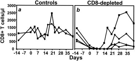
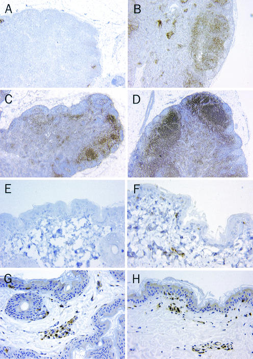
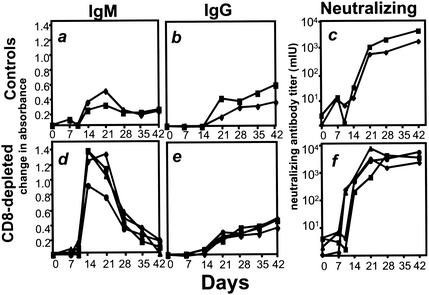
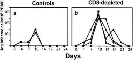
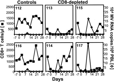
The creation of an improved vaccine for global measles control will require an understanding of the immune mechanisms of measles virus containment. To assess the role of CD8(+) cytotoxic T lymphocytes in measles virus clearance, rhesus monkeys were depleted of CD8(+) lymphocytes by monoclonal anti-CD8 antibody infusion and challenged with wild-type measles virus. The CD8(+) lymphocyte-depleted animals exhibited a more extensive rash, higher viral loads at the peak of virus replication, and a longer duration of viremia than did the control antibody-treated animals. These findings indicate a central role for CD8(+) lymphocytes in the control of measles virus infections and the importance of eliciting a cell-mediated immune response in new measles vaccine strategies.


Figures

FIG. 1.
FIG. 1.
CD8+ lymphocytes are depleted in the peripheral blood of rhesus monkeys as a result of intravenous injection of the mouse-human chimeric monoclonal anti-CD8 antibody cM-T807. The anti-CD8 antibody (four monkeys) or a control antibody (two monkeys) was administered intravenously on days −3, 0, and 4 of an MV infection. CD8+ T lymphocytes were quantitated by using a phycoerythrin-coupled monoclonal antibody that was able to bind to CD8 in the presence of cM-T807, as previously reported (28). When CD8+ T lymphocytes were not detectable, >95% of the remaining lymphocytes were CD20+ B cells or CD4+ T lymphocytes. (a) CD8+ T-lymphocyte counts in control antibody-treated monkeys. (b) CD8+ T-lymphocyte counts in monkeys treated with anti-CD8 antibody.
FIG. 2.
FIG. 2.
CD8+ lymphocytes are nearly eliminated in inguinal lymph nodes from CD8+ lymphocyte-depleted monkeys on day 7 after infection. CD8+ lymphocytes do not appear in the CD3+ lymphocytic infiltrate of the axillary measles rash on day 14 after infection. The formalin-fixed tissues were embedded in paraffin, sectioned at 5 μm, and immunostained either with polyclonal anti-CD3 antibodies (Dako, Carpinteria, Calif.) or with monoclonal anti-CD8 antibodies (1A5; Vector Laboratories, Burlingame, Calif.). Tissues stained for CD3 were preheated in a microwave oven for 20 min by using antigen unmasking solution (Vector Laboratories); those stained for CD8 were preheated in an electric pressure cooker for 15 min with Trilogy solution (Cell Marque Corp., Hot Springs, Ark.). For each primary antibody, an appropriate negative control was used at the same concentration: a rabbit immunoglobulin fraction for CD3 and mouse IgG1 for CD8. Sections were counterstained with hematoxylin. Lymph nodes from a monoclonal anti-CD8 antibody-treated monkey (A) and a control antibody-treated monkey (B) stained for expression of CD8 (magnification, ×20), lymph nodes from a monoclonal anti-CD8 antibody-treated monkey (C) and a control monkey (D) stained for expression of CD3 (magnification, ×20), axillary skin from a monoclonal anti-CD8 antibody-treated monkey (E) and a control antibody-treated monkey (F) stained for expression of CD8 (magnification, ×400), and axillary skin from a monoclonal anti-CD8 antibody-treated monkey (G) and a control antibody-treated monkey (H) stained for expression of CD3 (magnification, ×400) are shown.
FIG. 3.
FIG. 3.
Anti-MV antibody responses after MV infection in the CD8+ lymphocyte-depleted and control monkeys. Neutralizing antibody was measured in a plaque reduction assay using the Chicago-1 strain of MV and Vero cells as previously described (2). MV-specific IgM and IgG were measured in the MV IgG indirect enzyme immunoassay kit (Sigma) by substituting a horseradish-peroxidase-conjugated goat anti-monkey IgM (Nordic, Capistrano Beach, Calif.) and an alkaline phosphatase-conjugated rabbit anti-monkey IgG (Biomakor; Accurate Chemicals, Westbury, N.J.), respectively, for the secondary antibody. Sera were diluted 1:100, and secondary antibodies were diluted 1:2,000 in 1% normal rabbit serum and 0.05% Tween 20 in phosphate-buffered saline for all assays. Fast pNPP (Tab set-n2770; Sigma) was used as a substrate for the alkaline phosphatase-conjugated rabbit anti-monkey IgG, and plates were read by determining the optical density. Samples were tested in duplicate, and displayed values represent the mean change in absorbance from preinfected serum obtained from the same animal. (a) MV-specific IgM in control monkeys; (b) MV-specific IgG in control monkeys; (c) MV-neutralizing antibody in control monkeys; (d) MV-specific IgM in CD8-depleted monkeys; (e) MV-specific IgG in CD8-depleted monkeys; (f) MV-neutralizing antibodies in CD8-depleted monkeys.
FIG. 4.
FIG. 4.
Titers of infectious MV in the blood are greater in the CD8+ lymphocyte-depleted animals than in the control monkeys during early infection. Infectious virus was measured by cocultivation of PBL with B95-8 cells. (a) MV viremia in control monkeys; (b) MV viremia in CD8+ lymphocyte-depleted monkeys.
FIG. 5.
FIG. 5.
PBL MV RNA levels are higher in CD8+ lymphocyte-depleted than in control monkeys, and clearance coincides temporally with the repopulation of CD8+ lymphocytes. The CD8+ lymphocyte count (▪) and MV RNA level in PBL (⋄), measured by quantitative MV-specific RT-PCR, were assessed prospectively after MV infection in two control monkeys (animals 112 and 116) and four CD8+ lymphocyte-depleted monkeys (animals 113, 114, 115, and 117).

References

    1. Anonymous. 1999. Global measles control and regional elimination, 1998-1999. Morb. Mortal. Wkly. Rep. 48:1124-1130. - PubMed
    1. Auwaerter, P. G., P. A. Rota, W. R. Elkins, R. J. Adams, T. DeLozier, Y. Shi, W. J. Bellini, B. R. Murphy, and D. E. Griffin. 1999. Measles virus infection in rhesus macaques: altered immune responses and comparison of the virulence of six different virus strains. J. Infect. Dis. 180:950-958. - PubMed
    1. Burnet, F. M. 1968. Measles as an index of immunological function. Lancet ii:610-613. - PubMed
    1. Chen, R. T., L. E. Markowitz, P. Albrecht, J. A. Stewart, L. M. Mofenson, S. R. Preblud, and W. A. Orenstein. 1990. Measles antibody: reevaluation of protective titers. J. Infect. Dis. 162:1036-1042. - PubMed
    1. Forthal, D. N., G. Landucci, A. Habis, M. Zartarian, J. Katz, and J. G. Tilles. 1994. Measles virus-specific functional antibody responses and viremia during acute measles. J. Infect. Dis. 169:1377-1380. - PubMed

Publication types

MeSH terms