. 2003 Apr;77(7):4409-14.
doi: 10.1128/jvi.77.7.4409-4414.2003.

Disassembly of human immunodeficiency virus type 1 cores in vitro reveals association of Nef with the subviral ribonucleoprotein complex

Affiliations

Disassembly of human immunodeficiency virus type 1 cores in vitro reveals association of Nef with the subviral ribonucleoprotein complex

Brett M Forshey et al. J Virol. 2003 Apr.

Erratum in

  • J Virol. 2004 Apr;78(8):4384

Abstract

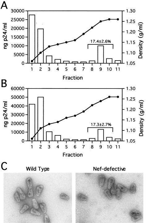
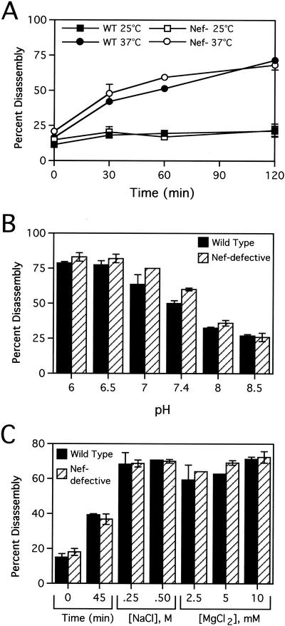
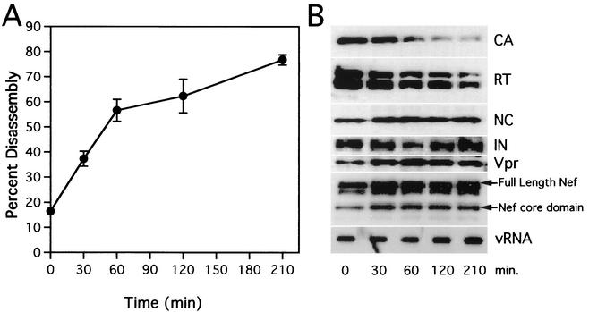
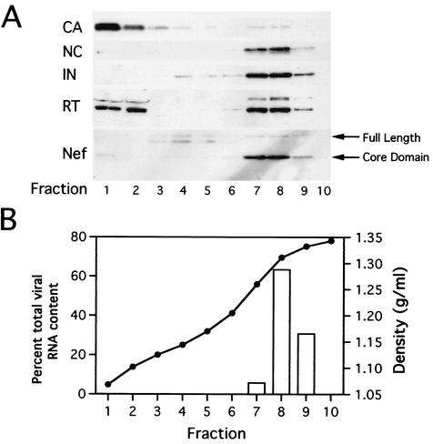
The human immunodeficiency virus type 1 (HIV-1) virulence factor Nef enhances viral infectivity in single-cycle infection assays and accelerates HIV-1 replication in vitro. It has been reported that the effects of Nef are mediated early after viral entry and before the completion of reverse transcription, as viral DNA synthesis is strongly attenuated during infection by Nef-defective virions. Our previous work has demonstrated that Nef is associated with mature HIV-1 cores, implicating Nef in the regulation of HIV-1 core stability. Here we report a comparative analysis of HIV-1 cores isolated from wild-type and Nef-defective particles. We observed no effect of Nef on HIV-1 core structure or stability; however, Nef cosedimented with a subviral ribonucleoprotein complex following dissociation of CA. These results indicate that Nef interacts tightly with an internal component of the HIV-1 core. They further suggest that virion-associated Nef may facilitate an early step in HIV-1 infection following dissociation of the viral capsid in the target cell.


Figures

FIG. 1.
FIG. 1.
Analysis of wild-type and Nef core yields and morphology. (A and B) Quantitation of wild-type and Nef-defective HIV-1 core yields. Concentrated virions were layered onto 30-to-70% (wt/vol) linear sucrose gradients atop a layer of Triton X-100. Following ultracentrifugation at 100,000 × g for 16 h at 4°C, fractions were collected from the top of the gradient and were analyzed by p24 ELISA. The density of each fraction was determined by refractometry. Panels A and B show the recovery of cores from wild-type and Nef-defective HIV-1 particles, respectively. Values shown represent the mean percentages (and standard deviations) of p24 found in the core-containing fractions for a minimum of five independent determinations. (C) Ultrastructural analysis of wild-type and Nef cores. Gradient fractions containing intact cores were pelleted at 100,000 × g for 30 min and resuspended in 15 μl of STE buffer. Samples were loaded onto carbon-coated grids and stained with 1% uranyl acetate. Samples were visualized at a magnification of ×40,000 in a Philips CM12 transmission electron microscope.
FIG. 2.
FIG. 2.
Nef does not detectably affect HIV-1 core disassembly in vitro. Purified wild-type (WT) and Nef-defective (Nef) cores were incubated in STE buffer at the indicated temperatures for the times shown (A) or at 37°C for 45 min at the indicated pH values (B) or NaCl or MgCl2 concentrations (C). Following incubation, the samples were subjected to ultracentrifugation at 100,000 × g for 15 min at 4°C. The extent of disassembly was determined as the percentage of the total CA protein in the reaction mixture present in the supernatant. Shown are mean values of duplicate determinations, with error bars representing the range of values.
FIG. 3.
FIG. 3.
Kinetic and biochemical analyses of HIV-1 core disassembly in vitro. Purified HIV-1 cores were incubated at 37°C for the indicated times, followed by separation of soluble and core-associated CA by ultracentrifugation. (A) Dissociation of CA from pelletable cores during incubation at 37°C. Supernatants and pellets were analyzed by p24 ELISA. The extent of disassembly was determined as the percentage of the total CA protein in the reaction mixture detected in the supernatant. Values represent means of triplicate determinations with error bars representing one standard deviation. (B) Biochemical analysis of particles remaining following disassembly of HIV-1 cores. Pellets from disassembly reactions shown in panel A were subjected to Western blotting and RNA slot blot analyses. The Western blot membrane was probed sequentially with rabbit anti-CA, anti-RT, anti-NC, anti-IN, anti-Vpr, and anti-Nef antibodies. RNA was extracted from pellets, denatured, and immobilized by vacuum filtration. Relative HIV-1 RNA levels were determined by hybridization with a 32P-labeled HIV-1 probe, followed by detection with autoradiography.
FIG. 4.
FIG. 4.
Gradient analysis of HIV-1 vRNPs isolated directly from virions. Concentrated virions were incubated in the presence of 0.5% Triton X-100 for 3 h at 37°C. Following incubation, the sample was loaded onto a 20-to-70% (wt/wt) sucrose gradient and subjected to ultracentrifugation at 100,000 × g for 16 h. Fractions were collected from the top of the gradient. (A) Proteins were precipitated with trichloroacetic acid and analyzed by Western blotting. (B) RNA was immobilized on nitrocellulose and viral RNA content was determined by hybridization with a 32P-labeled HIV-1 probe, followed by phosphorimager quantification. Fraction densities were determined by refractometric analysis of sucrose concentrations.

References

    1. Accola, M. A., A. Ohagen, and H. G. Gottlinger. 2000. Isolation of human immunodeficiency virus type 1 cores: retention of Vpr in the absence of p6gag. J. Virol. 74:6198-6202. - PMC - PubMed
    1. Ahmad, N., and S. Venkatesan. 1988. Nef protein of HIV-1 is a transcriptional repressor of HIV-1 LTR. Science 241:1481-1485. - PubMed
    1. Aiken, C. 1997. Pseudotyping human immunodeficiency virus type 1 (HIV-1) by the glycoprotein of vesicular stomatitis virus targets HIV-1 entry to an endocytic pathway and suppresses both the requirement for Nef and the sensitivity to cyclosporin A. J. Virol. 71:5871-5877. - PMC - PubMed
    1. Aiken, C., and D. Trono. 1995. Nef stimulates human immunodeficiency virus type 1 proviral DNA synthesis. J. Virol. 69:5048-5056. - PMC - PubMed
    1. Arora, V. K., B. L. Fredericksen, and J. V. Garcia. 2002. Nef: agent of cell subversion. Microbes Infect. 4:189-199. - PubMed

Publication types

MeSH terms

LinkOut - more resources