. 2003 Apr 1;100(7):4293-8.
doi: 10.1073/pnas.0730698100. Epub 2003 Mar 12.

Demonstration of functional coupling between gamma -aminobutyric acid (GABA) synthesis and vesicular GABA transport into synaptic vesicles

Affiliations

Demonstration of functional coupling between gamma -aminobutyric acid (GABA) synthesis and vesicular GABA transport into synaptic vesicles

Hong Jin et al. Proc Natl Acad Sci U S A. .

Abstract

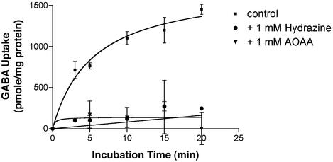
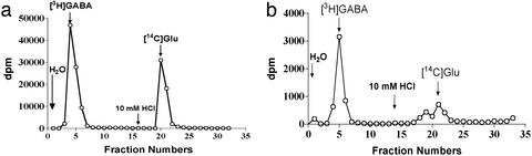
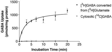
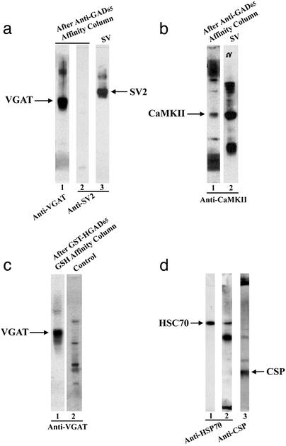
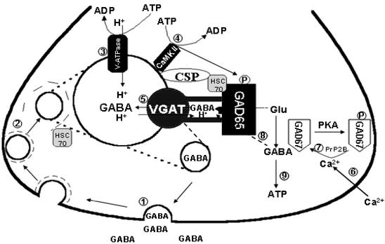
l-Glutamic acid decarboxylase (GAD) exists as both membrane-associated and soluble forms in the mammalian brain. Here, we propose that there is a functional and structural coupling between the synthesis of gamma-aminobutyric acid (GABA) by membrane-associated GAD and its packaging into synaptic vesicles (SVs) by vesicular GABA transporter (VGAT). This notion is supported by the following observations. First, newly synthesized [(3)H]GABA from [(3)H]l-glutamate by membrane-associated GAD is taken up preferentially over preexisting GABA by using immunoaffinity-purified GABAergic SVs. Second, the activity of SV-associated GAD and VGAT seems to be coupled because inhibition of GAD also decreases VGAT activity. Third, VGAT and SV-associated Ca(2+)calmodulin-dependent kinase II have been found to form a protein complex with GAD. A model is also proposed to link the neuronal stimulation to enhanced synthesis and packaging of GABA into SVs.


Figures

Figure 1
Figure 1
Isolation of specific GABAergic SVs by anti-GAD65-conjugated immunobeads. (a) Electron microscopic examination of SV preparations. (A) Control oxirane beads alone. (B and C) Anti-GAD65-coupled immunobeads coated with SVs. The dark dots associated with oxirane beads are SVs coupled to beads as indicated by the arrowheads. The bar in B indicates 1 μm, and the bar in C indicates 200 nm. (b) Immunoblotting test of various SV preparations with anti-VGAT. An amount of 20 μg of protein per lane was used. Lane 1, original SVs without treatment as control; lane 2, GABAergic SVs purified by anti-GAD65-conjugated immunobeads; lane 3, non-GABAergic SVs that were not retained by anti-GAD65 beads. Arrow indicates the position of VGAT.
Figure 2
Figure 2
Effect of GAD inhibitors on vesicular uptake of newly synthesized GABA. Uptake of GABA into SV was conducted as described in Materials and Methods under the following conditions: GABAergic SV alone (■), in the presence of the GAD inhibitor, 1 mM hydrazine (●) or 1 mM aminooxyacetate (▾). The uptake of GABA in the presence of the V-type ATPase inhibitor, bafilomycin A1 or in the absence of ATP was taken as nonspecific uptake, which was <5% of the total uptake. [3H]Glu was used as a substrate for the production of [3H]GABA. The specific uptake was obtained by subtracting the nonspecific uptake from the total uptake and was expressed as pmol per mg of protein. Data are presented as mean ± SD (n = 3).
Figure 3
Figure 3
Separation of GABA and glutamate on an anion exchange column. Separation of standard GABA (100,000 dpm) and Glu (60,000 dpm) as well as the extract of SV on an anion exchange column was conducted as described in Materials and Methods. (a) The elution pattern of standard GABA (first peak) and Glu (second peak) on an anion exchange column. (b) The elution pattern of the content extracted from SV (equivalent to 0.2 mg of protein, first peak) and standard [14C]Glu (second peak) on the same anion exchange column as in a. The first peak is identified as GABA because it has the same elution position as standard GABA in a.
Figure 4
Figure 4
A comparison of vesicular uptake of newly synthesized GABA and the preexisting GABA. [3H]Glu was used to generate newly synthesized [3H]GABA, whereas [14C]GABA was included as preexisting GABA in uptake experiments. GABA converted from Glu by SV-associated GAD was actively taken up (■), whereas the uptake of GABA preexisting in the solution was almost zero (▴). Data are presented as mean ± SD (n = 3).
Figure 5
Figure 5
Identification of VGAT, CaMKII, HSC70, and CSP as components of a GAD protein complex. (a) The GAD-associated protein complex in the solubilized SV preparation was purified by an anti-GAD65 affinity column as described in Materials and Methods. The anti-GAD65 immonoaffinity-purified protein complex (4.5 μg of protein per lane) was separated on SDS/PAGE, blotted on a nitrocellulose sheet, and probed with anti-VGAT (lane 1). The arrow indicates the position of VGAT. The same nitrocellulose membrane was stripped and reprobed with anti-SV2 antibodies (lane 2) showing no detectable SV2 in anti-GAD purified preparations. Lane 3 is SV alone as a control. The arrow indicates the position of SV2 protein, which is a marker for SV. (b) Lane 1 is the same as the lane 1 in Fig. 5a, except it was probed with anti-CaMKII showing staining at the position of CaMKII protein as indicated by the arrow. Lane 2 is SV alone as a control. (c) Immunoblotting analysis of the GAD protein complex formed between the solubilized SV solution and the recombinant GST-HGAD65 fusion protein probed with anti-VGAT. The arrow indicates the position of VGAT (lane 1). GST-GAD65 fusion protein alone showed no staining under the same conditions (lane 2). (d) Immunoblotting analysis of GABAergic SV (4.5 μg of protein per lane) isolated by anti-GAD65-conjugated beads with anti-HSC70 (lane 2). The arrow indicates the position of HSC70. Lane 1 is pure HSC70 alone as a marker. The same blot was stripped and restained with anti-CSP (lane 3). The arrow indicates the position of CSP.
Figure 6
Figure 6
A model depicting a structural and functional coupling between GABA synthesis and vesicular GABA transport into SV. GAD65 is anchored to SVs first through forming a protein complex with the chaperone protein, HSC70, followed by association of HSC70⋅GAD65 complex to CSP, VGAT, and CaMKII on SVs. The sequence of events leading from neuronal stimulation to activation of GAD65 and packaging of GABA into SVs is discussed in the text.

References

    1. Erlander M G, Tobin A J. Neurochem Res. 1991;16:215–226. - PubMed
    1. Dirkx R, Jr, Thomas A, Li L S, Lernmark Å, Sherwin R S, De Camilli P, Solimena M. J Biol Chem. 1995;270:2241–2246. - PubMed
    1. Sheikh S N, Martin D L. J Neurochem. 1996;66:2082–2090. - PubMed
    1. Kanaani J, Lissin D, Kash S F, Baekkekov S. J Biol Chem. 1999;274:37200–37209. - PubMed
    1. Solimena M, Aggujaro D, Muntzel C, Dirkx R, Jr, Butler M, De Camilli P, Hayday A. Proc Natl Acad Sci USA. 1993;90:3073–3077. - PMC - PubMed

Publication types

MeSH terms

LinkOut - more resources