. 2003 Mar;4(3):290-5.
doi: 10.1038/sj.embor.embor768.

The 37 kDa/67 kDa laminin receptor is required for PrP(Sc) propagation in scrapie-infected neuronal cells

Affiliations

The 37 kDa/67 kDa laminin receptor is required for PrP(Sc) propagation in scrapie-infected neuronal cells

Christoph Leucht et al. EMBO Rep. 2003 Mar.

Erratum in

  • EMBO Rep. 2003 Apr;4(4):439

Abstract

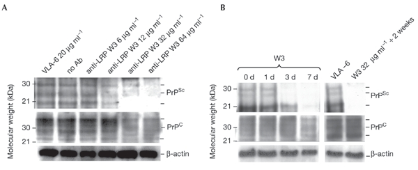
The accumulation of PrP(Sc) in scrapie-infected neuronal cells has been prevented by three approaches: (i) transfection of ScMNB cells with an antisense laminin receptor precursor (LRP) RNA-expression plasmid, (ii) transfection of ScN2a cells and ScGT1 cells with small interfering RNAs (siRNAs) specific for the LRP mRNA, and (iii) incubation of ScN2a cells with an anti-LRP/LR antibody. LRP antisense RNA and LRP siRNAs reduced LRP/LR expression and inhibited the accumulation of PrP(Sc) in these cells. The treatments also reduced PrP(c) levels. The anti-LRP/LR antibody, W3, abolished PrP(Sc) accumulation and reduced PrP(c) levels after seven days of incubation. Cells remained free of PrP(Sc) after being cultured for 14 additional days without the antibody, whereas the PrP(c) level was restored. Our results demonstrate the necessity of the laminin receptor (LRP/LR) for PrP(Sc) propagation in cultured cells and suggest that LRP/LR-specific antibodies could be used as powerful therapeutic tools in the treatment of transmissible spongiform encephalopathies.


Figures

Figure 1
Figure 1
Abolition of PrPSc propagation using laminin receptor precursor (LRP) antisense RNA. (A) Analysis by PCR with reverse transcription of total RNA extracts from transfected ScMNB cells. Oligodesoxythymidine-primed complementary DNA was amplified by PCR using specific primers for the pCI–neo plasmid. This gave a 322-bp cDNA fragment for the pCI–neo transfected cells and a 1,115-bp cDNA fragment for the pCI–neo–asLRP transfected cells. (B) A ribonuclease protection assay was carried out on total RNA from cells transfected with either pCI–neo or pCI–neo–asLRP; the RNA was then separated using a 5% acrylamide/urea gel. 5 μg or 10 μg of total RNA was used, and in both cases the level of LRP messenger RNA was reduced by 80–85% in cells transfected with pCI–neo–asLRP (quantified by posphorimaging). (C) Western blot analysis of cell lysates from pCI–neo- and pCI–neo–asLRP-transfected ScMNB cells assayed 48 hours after transfection. LRP was detected using the polyclonal anti-LRP/LR antibody, W3. β-actin was detected using an anti-β-actin antibody as loading control. (D) ScMNB cells were transfected with pCI–neo and pCI–neo–asLRP. The PrPSc content of ScMNB cells was analysed 72 h after transfection. The monoclonal anti-PrP antibody SAF70 was used for PrPSc detection and the SAF32 antibody was used for detection of PrPC.
Figure 2
Figure 2
Inhibition of PrPSc propagation using small interfering RNAs. (A) Western blot analysis of ScN2a cells transfected with small interfering RNAs (siRNAs). Cells were analysed 72 h after transfection using the polyclonal anti-laminin receptor (LR/LRP) antibody W3. (B) The effect of siRNAs on PrPSc propagation was assayed 72 h after transfection (left panel). The time-dependent effect of siRNA-LRP3 on PrPSc propagation (right panel) was analysed using the SAF70 antibody; PrPC was detected with the SAF32 antibody. β-actin was detected using an anti-β-actin antibody as a loading control (A, B). (C) Western blot analysis of siRNA-transfected ScGT1 cells at 72 h after transfection. The cells were analysed using the monoclonal antibodies LR43512 (lower panel) and SAF84 (upper panel). All samples were normalized to equal protein concentrations.
Figure 3
Figure 3
The effect of the W3 anti-laminin receptor (LRP/LR) antibody on PrPSc propagation. (A) ScN2a cells were incubated with W3 at varying concentrations. The PrPSc content was determined after a 72 h incubation with W3. An anti VLA-6 (integrin-type laminin receptor) antibody was used as a control. PrPSc was detected using the A7 polyconal antibody; PrPC was detected with the SAF32 antibody. (B) ScN2a cells were incubated with W3 at 32 μg ml−1 for varying durations. The last lane shows W3-treated ScN2a cells after an additional 2-week incubation without any antibody. PrPSc was detected with the SAF 70 antibody, PrPc was detected with the SAF32 antibody. β-actin was detected using an anti-β-actin antibody as a loading control.

References

    1. Aguzzi A. & Weissmann C. (1998) Prion diseases. Haemophilia, 4, 619–627. - PubMed
    1. Bosque P.J. & Prusiner S.B. (2000) Cultured cell sublines highly susceptible to prion infection. J. Virol., 74, 4377–4386. - PMC - PubMed
    1. Caughey B. & Raymond G.J. (1991) The scrapie-associated form of PrP is made from a cell surface precursor that is both protease- and phospholipasesensitive. J. Biol. Chem., 266, 18217–18223. - PubMed
    1. Douville P.J. & Carbonetto S. (1992) Genetic linkage analysis in recombinant inbred mice of P40, a putative clone for the high-affinity laminin receptor. Mamm. Genome, 3, 438–446. - PubMed
    1. Elbashir S.M., Harborth J., Lendeckel W., Yalcin A., Weber K. & Tuschl T. (2001) Duplexes of 21-nucleotide RNAs mediate RNA interference in cultured mammalian cells. Nature, 411, 494–498. - PubMed

Publication types