

Parkin binds the Rpn10 subunit of 26S proteasomes through its ubiquitin-like domain
- PMID: 12634850
- PMCID: PMC1315892
- DOI: 10.1038/sj.embor.embor764
Parkin binds the Rpn10 subunit of 26S proteasomes through its ubiquitin-like domain
Abstract
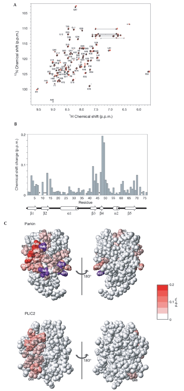
Parkin, a product of the causative gene of autosomal-recessive juvenile parkinsonism (AR-JP), is a RING-type E3 ubiquitin ligase and has an amino-terminal ubiquitin-like (Ubl) domain. Although a single mutation that causes an Arg to Pro substitution at position 42 of the Ubl domain (the Arg 42 mutation) has been identified in AR-JP patients, the function of this domain is not clear. In this study, we determined the three-dimensional structure of the Ubl domain of parkin by NMR, in particular by extensive use of backbone (15)N-(1)H residual dipolar-coupling data. Inspection of chemical-shift-perturbation data showed that the parkin Ubl domain binds the Rpn10 subunit of 26S proteasomes via the region of parkin that includes position 42. Our findings suggest that the Arg 42 mutation induces a conformational change in the Rpn10-binding site of Ubl, resulting in impaired proteasomal binding of parkin, which could be the cause of AR-JP.
Figures

References
-
- Beal R.E., Toscano-Cantaffa D., Young P., Rechsteiner M. & Pickart C.M. (1998) The hydrophobic effect contributes to polyubiquitin chain recognition. Biochemistry, 37, 2925–2934. - PubMed
-
- Brünger A.T. et al. . (1998) Crystallography & NMR system: A new software suite for macromolecular structure determination. Acta Crystallogr. D Biol. Crystallogr., 54, 905–921. - PubMed
-
- Buchberger A. (2002) From UBA to UBX: new words in the ubiquitin vocabulary. Trends Cell Biol., 12, 216–221. - PubMed
-
- Chung K.K. et al. . (2001) Parkin ubiquitinates the alphasynuclein-interacting protein, synphilin-1: implications for Lewy-body formation in Parkinson disease. Nature Med., 7, 1144–1150. - PubMed
-
- Clore G.M., Gronenborn A.M. & Bax A. (1998) A robust method for determining the magnitude of the fully asymmetric alignment tensor of oriented macromolecules in the absence of structural information. J. Magn. Reson., 133, 216–221. - PubMed
Publication types
MeSH terms
Substances
Associated data
- Actions
LinkOut - more resources
Full Text Sources
Other Literature Sources
Molecular Biology Databases
Research Materials
