. 2003 Feb 27;2(1):3.
doi: 10.1186/1475-2883-2-3.

Animated documentation of the filaria dance sign (FDS) in bancroftian filariasis

Affiliations

Animated documentation of the filaria dance sign (FDS) in bancroftian filariasis

Sabine Mand et al. Filaria J. .

Abstract

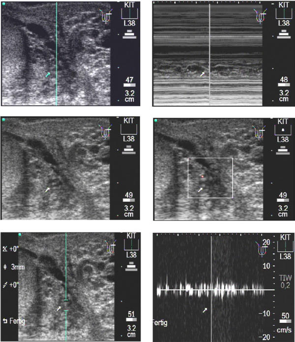
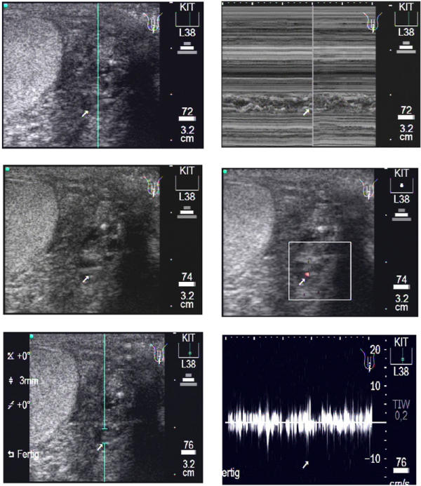
BACKGROUND: Ultrasonography is able to detect adult Wuchereria bancrofti worms in scrotal lymphatic vessels of infected men on account of the characteristic pattern of adult worm movements, known as the filarial dance sign. Furthermore, the technique is able to delineate associated pathology, such as hydrocoele and lymphoedema, which can be diagnosed in early stages. Ultrasonography is also useful in the assessment of macrofilaricidal effects of antifilarial medication.The purpose of this study was to evaluate the usefulness of scrotal ultrasonography, in combination with a new method of digital documentation, in men infected with Wuchereria bancrofti. METHODS: Ultrasonography of the scrotal areas was carried out in 33 male patients from an endemic area in Ghana using a hand-carried ultrasound system and a linear array transducer at 7.5 MHz. Wuchereria bancrofti infection was also assessed by quantification of night blood microfilaraemia and semi-quantitative detection of circulating filarial antigen. Ultrasound findings were documented by print outs and by Digital Video sequences directly exported from the ultrasound machine which were edited in Final Cut Pro 3ledR; and exported, using QuickTimecircledR; Pro, as MPEG-1 video. RESULTS: Worm nests, i.e. dilated lymphatic vessels with the characteristic movement patterns of worms, were found in all patients, and typical examples of larger as well as smaller nests are presented through MPEG-1 video in b- and m-modes as well as Colour Doppler and Pulse Wave Doppler images. CONCLUSION: In this study, the filarial dance sign is being made available on the Internet to readers through MPEG-1 video. This method allows for demonstration of movement patterns rather than static images. In addition, the pathologic ultrasonographic signs of filariasis can be rapidly relayed over great distances and may be helpful to other investigators or clinicians in the diagnosis of patients infected with Wuchereria bancrofti.


Figures

Figure 1
Figure 1
Transverse scan of a patient's left testis. Upper left: In para-testicular position an enlarged lymphatic vessel with one or more adult worms can be seen (arrow). The blue line depicts the cursor position of the following m-mode, upper right. The corresponding video image can be seen as Additional File 1: Movie1A.mpg. Middle left: The same worm nest as seen above. Middle right: The use of the Colour Power Doppler shows that this worms nests contains much free lymphatic fluid which induces a red signal due to the worm movements in different parts of the worm nest. Different to vessels, the signal is non-rhythmic and non-pulsating. The corresponding video image can be seen as Additional File 2: Movie1B.mpg. Lower left: The same worm nests as seen above. The caliper marks the position at which the Pulse Wave Doppler (PWD) is performed. Lower right: After switch to the PWD mode the filaria dance sign (FDS) is seen as an undulating band as a function of time, with sharp, irregular peaks (turquoise arrowhead). The corresponding video image can be seen as Additional File 3: Movie1C.mpg.
Figure 2
Figure 2
Transverse scan of a patient's left testis. Upper left: The b-mode image shows the worm nest (arrow) in para-testicular location with the cursor (blue line) positioned for the following m-mode image. Upper right: In the m-mode image the body wall (arrow) of the adult worms is detectable as double layers in form of wavy bands. The corresponding video image can be seen as Additional File 4: Movie2A.mpg. Middle left: The same worm nests as above. In contrast to the worm nest seen in fig. 1 this worm nest is smaller and contains less lymphatic fluid. Middle right: The Colour power Doppler-mode shows clearly less red signals than in Fig. 1 (middle right, or movie 1B). The corresponding video image can be seen as Additional File 5: Movie2B.mpg. Lower left: The same worm nests as above. The caliper for the PWD is positioned where the worm nest is located. Lower right: The following PWD confirms the fact that this worm nest is smaller than the one Fig. 1, as documented by lower peaks, again characterized by the irregular undulating band in contrast to pulsating amplitudes caused by arteries. The corresponding video image can be seen as Additional File 6: Movie2C.mpg.
Figure 3
Figure 3
Longitudinal scan of a patient's left testis. Upper left: A medium sized para-testicular worm nest (arrow) is presented in this b-mode image. Upper right: The following m-mode section is positioned at the location of the largest diameter of the worm nest. The corresponding video image can be seen as Additional File 7: Movie3A.mpg. Middle left: The same worm nest as above. Middle right: The Colour Doppler-mode presents very few red signals as sign of less lymphatic fluid moved by the adult worms in this dilated lymphatic vessel. The corresponding video image can be seen as Additional File 8: Movie3B.mpg. Lower left: The same worm nest as above. The caliper for the PWD is positioned where the worm nest is located. Lower right: The Pulse Wave Doppler-mode confirms the medium sized worm nest by the irregular undulating band caused by the typical movements of the adult worms. The corresponding video image can be seen as Additional File 9: Movie3C.mpg.

References

    1. Ottesen EA. The Global Programme to Eliminate Lymphatic Filariasis. Trop Med Int Hlth. 2000;5:591–594. doi: 10.1046/j.1365-3156.2000.00620.x. - DOI - PubMed
    1. Anonymous The Global Alliance for the Elimination of Lymphatic Filariasis - Epidemiology. http://www.filariasis.org/ 2002.
    1. Amaral F, Dreyer G, Figueredo-Silva J, Norões J, Cavalcanti A, Samico SC, Santos A, Coutinho A. Live adult worms detected by ultrasonography in human Bancroftian filariasis. Am J Trop Med Hyg. 1994;50:753–757. - PubMed
    1. Dreyer G, Amaral F, Norões J, Medeiros Z. Ultrasonographic evidence for stability of adult worm location in bancroftian filariasis. Trans R Soc Trop Med Hyg. 1994;88:558. - PubMed
    1. Dreyer G, Addiss D, Roberts J, Norões J. Progression of lymphatic vessel dilatation in the presence of living adult Wuchereria bancrofti. Trans R Soc Trop Med Hyg. 2002;96:157–161. - PubMed

LinkOut - more resources