

Cognitive impairment in children with hemoglobin SS sickle cell disease: relationship to MR imaging findings and hematocrit
- PMID: 12637286
- PMCID: PMC7973593
Cognitive impairment in children with hemoglobin SS sickle cell disease: relationship to MR imaging findings and hematocrit
Abstract
Background and purpose: Children with hemoglobin SS sickle cell disease are known to suffer cognitive impairment if they have silent infarct, but recent evidence suggests that patients with hemoglobin SS sickle cell disease may be impaired even if they are free of infarction. We test a hypothesis that cognitive impairment in children with hemoglobin SS sickle cell disease is associated with low hematocrit and MR imaging abnormalities.
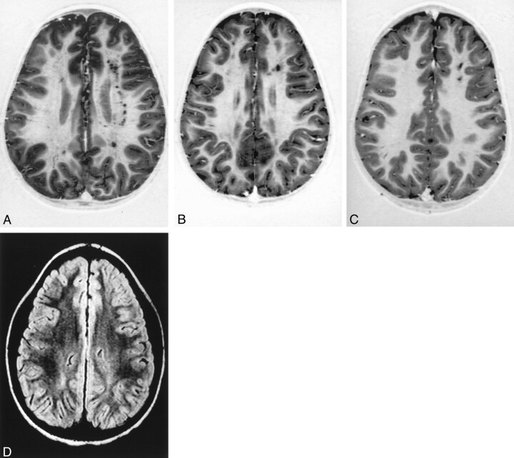
Methods: A cohort of 49 patients was examined, all of whom had hemoglobin SS sickle cell disease but no history of clinical stroke. The Wechsler scales, which are standardized and age-adjusted, were used to assess cognitive function. Patients also underwent MR imaging examination of the brain, and hematocrit was measured in a subset of 45 patients. MR images were evaluated by at least two readers, and abnormal imaging findings were evaluated by at least three readers. Any lesion was sufficient to be classified as abnormal, with lesions defined to include lacunar infarction, encephalomalacia, or leukoencephalopathy. Hematocrit data were used if obtained within 3 months of psychometric testing and if there were no confounding events in the patients' charts. Wechsler test scores were then evaluated in relation to imaging findings and hematocrit values.
Results: Patients with imaging abnormalities had more cognitive impairment than did patients with normal imaging findings in verbal intelligence quotient (P <.02) and verbal comprehension (P <.01). Patients with low hematocrit had cognitive impairment shown by many performance measures, including full-scale intelligence quotient (P <.006), verbal comprehension (P <.006), and freedom from distractibility (P <.02). Multivariate analysis showed that MR imaging and hematocrit were independent predictors of full-scale intelligence quotient.
Conclusion: Focal brain injury, revealed by MR imaging, is associated with cognitive impairment, but our data suggest that diffuse brain injury may also contribute to impairment. These findings show that impairment is multifactorial and suggest that chronic brain hypoxia is part of the pathophysiology of sickle cell disease.
Figures
References
-
- DeBaun MR, Schatz J, Siegel MJ, et al. Cognitive screening examinations for silent cerebral infarctions in sickle cell disease. Neurology 1998;50:1678–1682 - PubMed
-
- Steen RG, Xiong X, Mulhern RK, Langston JW, Wang WC. Subtle brain abnormalities in children with sickle cell disease: relationship to blood hematocrit. Ann Neurol 1999;45:279–286 - PubMed
-
- Brown RT, Buchannan I, Doepke K, et al. Cognitive and academic functioning in children with sickle cell disease. J Clin Child Psychol 1993;22:207–218
-
- Steen RG, Xiong X, Mulhern RK, Langston JW, Wang WC. Reply to Letter to the Editor. Ann Neurol 2000;47:280
-
- Wechsler D. Wechsler Intelligence Scale for Children. 3rd ed. San Antonio: The Psychological Corp.; 1991
Publication types
MeSH terms
Grants and funding
LinkOut - more resources
Full Text Sources
Medical