

Subtraction helical CT angiography of intra- and extracranial vessels: technical considerations and preliminary experience
- PMID: 12637296
- PMCID: PMC7973630
Subtraction helical CT angiography of intra- and extracranial vessels: technical considerations and preliminary experience
Abstract
Background and purpose: Reported CT angiographic (CTA) subtraction methods are not simple, robust, or real time. We investigated a novel technique for semiautomated digital subtraction CTA of the intracranial and extracranial arteries.
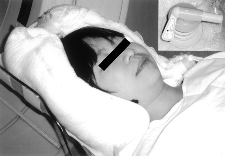
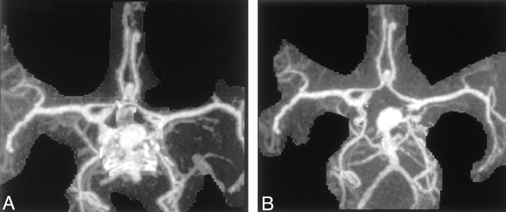
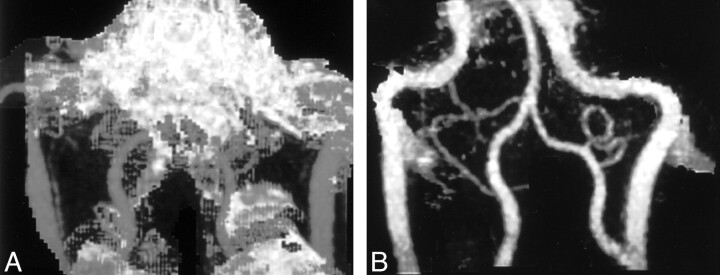
Methods: Thirty patients underwent precontrast (low milliampere-seconds) and postcontrast (pitch, 1.5; collimation, 1-2.5 mm) helical imaging with a vacuum-type head holder to facilitate image registration and minimize movement. A reconstructed three-dimensional model of the precontrast bone dataset was subtracted from the postcontrast dataset to produce subtracted maximum-intensity-projection angiograms. Experienced (operator 1) and less-experienced (operator 2) staff performed the standard and subtraction reconstructions, and image generation time and quality (graded 1-5) were compared. A third operator blinded to the method assessed the hard-copy image quality.
Results: Image quality with subtraction postprocessing was significantly better with both operators (operator 1, mean improvement of 0.87 grade, median improvement of 1 grade, P <.001; operator 2, mean improvement of 0.63 grade, median improvement of 1 grade, P <.001). Hard-copy image quality was better with the subtraction method (operator 1, P >.001; operator 2, P <.001). Blood vessels at the base of the brain were better demonstrated on subtraction images in 13 of 14 examinations. For the less experienced operator, the reconstruction time was significantly less with the subtraction method than with the conventional method (mean, 7.5 vs 10.1 minutes; P =.001).
Conclusion: When separation of the vasculature from bone is important and technically difficult, digital subtraction CTA offers a potential advantage. This semiautomated technique is fast and easy to learn, and variably experienced staff can use it.
Figures
Comment in
-
Subtraction helical CT angiography of intra- and extracranial vessels: technical considerations and preliminary experience--rediscovery of matched mask bone elimination?AJNR Am J Neuroradiol. 2003 Aug;24(7):1491; author reply 1491-2. AJNR Am J Neuroradiol. 2003. PMID: 12917153 Free PMC article. No abstract available.
Similar articles
-
Subtraction helical CT angiography of intra- and extracranial vessels: technical considerations and preliminary experience--rediscovery of matched mask bone elimination?AJNR Am J Neuroradiol. 2003 Aug;24(7):1491; author reply 1491-2. AJNR Am J Neuroradiol. 2003. PMID: 12917153 Free PMC article. No abstract available.
-
Prospective evaluation of carotid artery stenosis: elliptic centric contrast-enhanced MR angiography and spiral CT angiography compared with digital subtraction angiography.AJNR Am J Neuroradiol. 2003 May;24(5):1012-9. AJNR Am J Neuroradiol. 2003. PMID: 12748115 Free PMC article.
-
[Dual-energy CT angiography for evaluation of internal carotid artery stenosis and occlusion].Zhongguo Yi Xue Ke Xue Yuan Xue Bao. 2009 Apr;31(2):215-20. Zhongguo Yi Xue Ke Xue Yuan Xue Bao. 2009. PMID: 19507603 Chinese.
-
Bone-subtraction CT angiography for the evaluation of intracranial aneurysms.AJNR Am J Neuroradiol. 2006 Jan;27(1):55-9. AJNR Am J Neuroradiol. 2006. PMID: 16418356 Free PMC article.
-
[Dolichoectatic intracranial arteries. Advances in images and therapeutics].Medicina (B Aires). 1995;55(1):59-68. Medicina (B Aires). 1995. PMID: 7565039 Review. Spanish.
Cited by
-
Relationship between encephalopathy and portal vein-vena cava shunt: value of computed tomography during arterial portography.World J Gastroenterol. 2004 Jul 1;10(13):1939-42. doi: 10.3748/wjg.v10.i13.1939. World J Gastroenterol. 2004. PMID: 15222041 Free PMC article. Clinical Trial.
-
Dual-energy direct bone removal CT angiography for evaluation of intracranial aneurysm or stenosis: comparison with conventional digital subtraction angiography.Eur Radiol. 2009 Apr;19(4):1019-24. doi: 10.1007/s00330-008-1213-5. Epub 2008 Nov 11. Eur Radiol. 2009. PMID: 19002466
-
Clinical evaluation of bone-subtraction CT angiography (BSCTA) in head and neck imaging.Eur Radiol. 2006 Apr;16(4):889-97. doi: 10.1007/s0330-005-0032-1. Epub 2005 Nov 3. Eur Radiol. 2006. PMID: 16267665
-
Subtraction helical CT angiography of intra- and extracranial vessels: technical considerations and preliminary experience--rediscovery of matched mask bone elimination?AJNR Am J Neuroradiol. 2003 Aug;24(7):1491; author reply 1491-2. AJNR Am J Neuroradiol. 2003. PMID: 12917153 Free PMC article. No abstract available.
-
Dual energy bone subtraction in computed tomography angiography of extracranial-intracranial bypass: feasibility and limitations.Eur Radiol. 2011 Apr;21(4):750-6. doi: 10.1007/s00330-010-1973-6. Epub 2010 Oct 4. Eur Radiol. 2011. PMID: 20890761
References
-
- Young N, Dorsch NW, Kingston RJ, et al. Spiral CT scanning in the evaluation of the Circle of Willis. Surg Neurol 1998;50:50–60 - PubMed
-
- White PM, Wardlaw JM, Easton V. Can non-invasive imaging tests accurately detects intracranial aneurysms? A systematic review. Radiology 2000;217:361–370 - PubMed
-
- Sameshima T, Futami S, Morita Y, et al. Clinical usefulness and problems with three dimensional CT angiography for the evaluation of atherosclerotic stenosis of the carotid artery: comparison with conventional angiography, MRA and ultrasound sonography. Surg Neurol 1999;51:300–309 - PubMed
-
- Schwartz RB, Tice HM, Hooten SM, et al. Evaluation of cerebral aneurysms with helical CT: correlation with conventional angiography and MR angiography. Radiology 1994;192:717–722 - PubMed
MeSH terms
LinkOut - more resources
Full Text Sources
Medical