

Review
CT and MR imaging of intracerebral amyloidoma: case report and review of the literature
Affiliations
- PMID: 12637308
- PMCID: PMC7973613
Item in Clipboard
Review
CT and MR imaging of intracerebral amyloidoma: case report and review of the literature
AJNR Am J Neuroradiol.
2003 Mar.
Abstract
Intracerebral amyloidoma is the least common form of amyloid deposition in the brain. CT and MR imaging features in a case of pathologically proved cerebral amyloidoma are presented, and the available literature is reviewed. Typical imaging features of this entity include solitary or multiple supratentorial white matter masses that are hyperattenuated on nonenhanced CT. They have little or no mass effect on surrounding structures, extend medially up to the lateral ventricle wall, and have fine, irregular, enhancing margins.
Figures
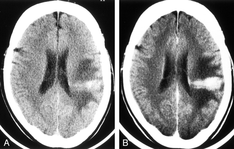
CT scans obtained in 54-year-old woman with intracerebral amyloidoma. A, Axial non-contrast enhanced CT scan of the brain reveals a hyperattenuated comet-shaped mass in the left posterior frontal white matter. Significant vasogenic edema is also present around the mass. B, The mass enhances after administration of contrast material. Note the medial extension of the mass up to the lateral ventricular ependyma.
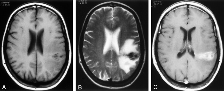
MR images obtained in 54-year-old woman with intracerebral amyloidoma. A, The mass is hypointense on this axial T1-weighted image (551/5/2 [TR/TE/NEX]). B, The mass is hypointense on this axial T2-weighted image (4110/110/2). C, The mass is hyperintense on this T1-weighted postcontrast axial image.
References
-
- Glenner GG. Amyloid deposits and amyloidosis. The β-fibrillosis. N Engl J Med 1980;302:1283–92, 1333–43 - PubMed
-
- Eriksson L, Sletten K, Benson L, Westermark P. Tumor-like localized amyloid of the brain is derived from immunoglobin light chain. Scand J Immunol 1993;37:623–626 - PubMed
-
- Caerts B, Mol V, Sainte T, Wilms G, Van Den Bergh V, Stessens L. CT and MRI of amyloidoma of the CNS. Eur Radiol 1997;7:474–476 - PubMed
-
- Salytkov S. Zur Frage des lokalen Amyloids des Hirengefabe. Bemerkung Zu dem Aufsatz von morgenstern Virchows Arch [A] 1935;295:590
Publication types
MeSH terms
LinkOut - more resources
Full Text Sources
Medical