. 2003 Mar;111(6):843-50.
doi: 10.1172/JCI16502.

In vivo derivation of glucose-competent pancreatic endocrine cells from bone marrow without evidence of cell fusion

Affiliations

In vivo derivation of glucose-competent pancreatic endocrine cells from bone marrow without evidence of cell fusion

Andreea Ianus et al. J Clin Invest. 2003 Mar.

Abstract

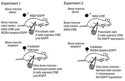
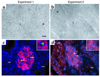
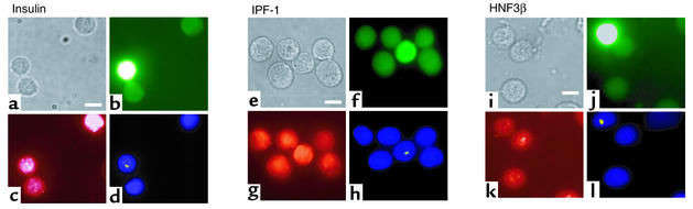
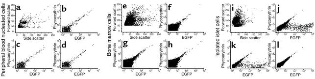
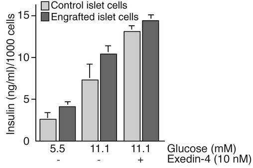
Bone marrow harbors cells that have the capacity to differentiate into cells of nonhematopoietic tissues of neuronal, endothelial, epithelial, and muscular phenotype. Here we demonstrate that bone marrow-derived cells populate pancreatic islets of Langerhans. Bone marrow cells from male mice that express, using a CRE-LoxP system, an enhanced green fluorescent protein (EGFP) if the insulin gene is actively transcribed were transplanted into lethally irradiated recipient female mice. Four to six weeks after transplantation, recipient mice revealed Y chromosome and EGFP double-positive cells in their pancreatic islets. Neither bone marrow cells nor circulating peripheral blood nucleated cells of donor or recipient mice had any detectable EGFP. EGFP-positive cells purified from islets express insulin, glucose transporter 2 (GLUT2), and transcription factors typically found in pancreatic beta cells. Furthermore, in vitro these bone marrow-derived cells exhibit - as do pancreatic beta cells - glucose-dependent and incretin-enhanced insulin secretion. These results indicate that bone marrow harbors cells that have the capacity to differentiate into functionally competent pancreatic endocrine beta cells and that represent a source for cell-based treatment of diabetes mellitus. The results generated with the CRE-LoxP system also suggest that in vivo cell fusion is an unlikely explanation for the "transdifferentiation" of bone marrow-derived cells into differentiated cell phenotypes.


Figures

Figure 1
Figure 1
Mouse bone marrow transplantation protocol. Experiment 1: Bone marrow from male INS2*EGFP mice was injected into irradiated female wild-type mice. Experiment 2: Bone marrow from male INS2-CRE mice was injected into irradiated female ROSA-stoplox-EGFP mice.
Figure 2
Figure 2
Immunofluorescence and FISH for Y chromosome of pancreatic 5- to 7-μm frozen sections from recipient mice of experiments 1 and 2 (see Figure 1). (a and b) Bright-field phase images. (c and d) Composite overlay image of immunofluorescence for insulin (red) and EGFP (green), and FISH for Y chromosome (yellow) and DAPI staining of nuclei (blue). Y chromosome–positive cells (arrows) are present in the islet (i) and in the exocrine portions of the pancreas (e). EGFP is present only in insulin-positive cells in the islet. Scale bar, 10 μm; ×400. Insets in c and d are magnified areas of EGFP and insulin double-positive cells containing a Y chromosome.
Figure 3
Figure 3
Immunofluorescence and FISH of isolated, dispersed pancreatic islet cells from experiment 1 (see Figure 1). Images of identical fields are shown in the respective panels: (ad) insulin, (eh) IPF-1, and (il) HNF3β immunodetection. (a, e, and i)Bright-field phase; (b, f, and j) EGFP (note slight autofluorescence of control isolated islet cells); (c, g, and k) Immunostaining with rhodamine X–labeled secondary antibody for panels. (c) Insulin, (g) IPF-1, (k) HNF3β, and (d h, and l) FISH for Y chromosome (in yellow) and nucleus stain with DAPI (blue). Y chromosome is present only in EGFP-positive cells. Scale bar, 5 μm; ×630.
Figure 4
Figure 4
Cell sorting and fluorescence analysis. (ad) Peripheral blood nucleated cells; (eh) bone marrow cells; (il) isolated and dispersed islet cells. (a, e, and i) Representational scatter plot of side (x axis) and forward (y axis) scatter of respective cells. (bd, fh, and jl) Fluorescence scans (x axis, EGFP; y axis, phycoerythrin [red] filter) of respective cells. (b, f, and j) Cells from INS*EGFP donor mice (experiment 1). (c, g, and k) Cells from irradiated wild-type mice transplanted with bone marrow from INS*EGFP mice (experiment 1). (d, h, and l) Cells from irradiated ROSA-stoplox-EGFP mice transplanted with bone marrow from INS2-CRE mice (experiment 2). No fluorescence is detectable in donor or recipient peripheral blood nucleated cells or bone marrow cells. EGFP is detectable in islets of donor animals of experiment 1 and in the recipients of experiment 1. In bd, fh, and jl, cells in the line equidistant from both axes show some autofluorescence, and cells in the lower right quadrant have EGFP signal. See Figure 1 for description of experiments 1 and 2.
Figure 5
Figure 5
RT-PCR of isolated and FACS-sorted cells from experiment 1. (a) Peripheral blood nucleated cells (PBNCs) of recipient mice express cyclophilin (lane 1) as well as CD45 (lane 2), and EGFP-expressing cells derived from pancreatic islets express cyclophilin (lane 3), lack CD45 (lane 4), and express insulin (lane 5). (b) EGFP-expressing cells isolated from pancreatic islets do not express the stem cell marker oct-3/4. Lane 1: Positive control oct-3/4 plasmid. Bone marrow cells express cyclophilin (lane 2) and oct-3/4 (lane 3), and EGFP-expressing cells from pancreatic islets express cyclophilin (lane 4) but not oct-3/4 (lane 5). (c) EGFP-expressing cells isolated from pancreatic islets express cyclophilin (lane 1), insulin I (lane 2), IPF-1 (lane 4), and HNF3β (lane 5). Lane 3 is empty. (d) EGFP-expressing cells derived from pancreatic islets express cyclophilin (lane 1), insulin II (lane 2), insulin I (lane 3), GLUT2 (lane 4), HNF1α (lane 5), HNF1β (lane 6), and PAX6 (lane 7).
Figure 6
Figure 6
Insulin secretion of EGFP-positive cells isolated from islets is similar to insulin secretion of control islet cells. Insulin secretion after glucose or glucose and exendin-4 stimulation of isolated EGFP-positive cells from experiment 1 is shown as dark gray bars. Control cells were isolated from an INS*EGFP mouse and treated identically (light gray bars). One thousand dispersed islet cells were collected for EGFP expression by FACS and/or manual picking, cultured for 24 hours at 5.5 mM glucose or 11.1 mM glucose for 10 hours. Additional cells were incubated in 11.1 mM glucose for 10 hours and then with 10 nM exendin-4 for 4 more hours. Supernatant was collected for insulin measurements (ELISA). Results of three separate assays each performed in duplicate are shown as mean ± SD.

Comment in

References

    1. Rosenberg L, Vinik AI, Pittenger GL, Rafaeloff R, Duguid WP. Islet-cell regeneration in the diabetic hamster pancreas with restoration of normoglycaemia can be induced by a local growth factor(s) Diabetologia. 1996;39:256–262. - PubMed
    1. Rafaeloff R, Barlow SW, Rosenberg L, Vinik AI. Expression of Reg gene in the Syrian golden hamster pancreatic islet regeneration model. Diabetologia. 1995;38:906–913. - PubMed
    1. Stoffers DA, et al. Insulinotropic glucagon-like peptide 1 agonists stimulate expression of homeodomain protein IDX-1 and increase islet size in mouse pancreas. Diabetes. 2000;49:741–748. - PubMed
    1. Guz Y, Nasir I, Teitelman G. Regeneration of pancreatic beta cells from intra-islet precursor cells in an experimental model of diabetes. Endocrinology. 2001;142:4956–4968. - PubMed
    1. Fernandes A, et al. Differentiation of new insulin-producing cells is induced by injury in adult pancreatic islets. Endocrinology. 1997;138:1750–1762. - PubMed

Publication types