. 2003 Mar;111(6):851-7.
doi: 10.1172/JCI16584.

Acceleration of type 1 diabetes mellitus in proinsulin 2-deficient NOD mice

Affiliations

Acceleration of type 1 diabetes mellitus in proinsulin 2-deficient NOD mice

Karine Thébault-Baumont et al. J Clin Invest. 2003 Mar.

Abstract

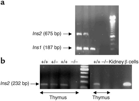
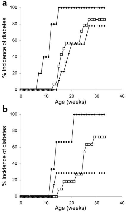
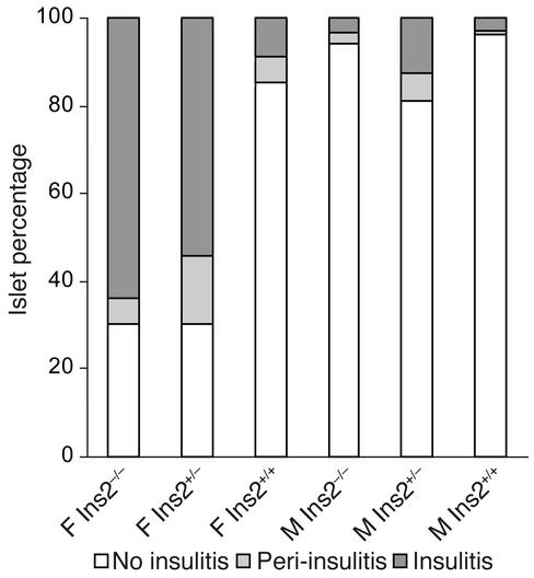
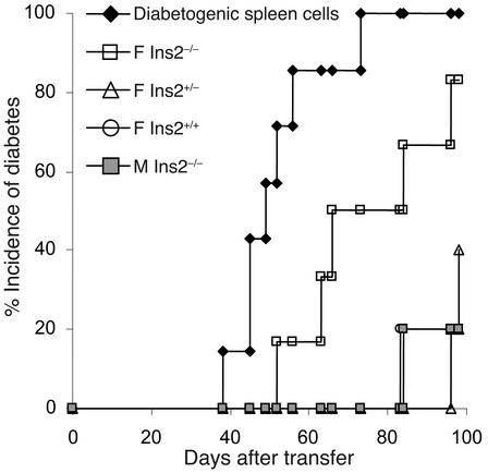
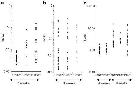
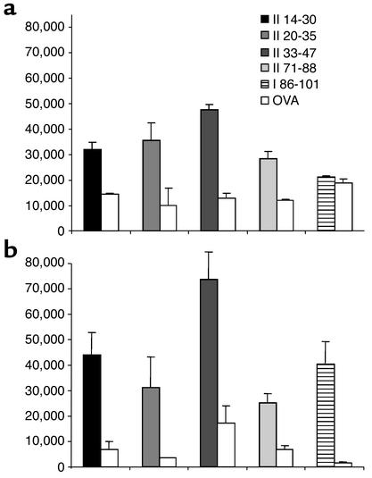
Accumulating evidence favors a role for proinsulin as a key autoantigen in diabetes. In the mouse, two proinsulin isoforms coexist. Most studies point to proinsulin 2 as the major isoform recognized by T cells in the NOD mouse. We studied mice in which a null proinsulin 2 mutation was transferred from proinsulin 2-deficient 129 mice onto the NOD background along with 16 genetic markers (including I-A(g7) MHC molecule) associated with diabetes. Intercross mice from the fourth backcross generation showed that proinsulin 2(-/-) mice develop accelerated insulitis and diabetes. The high prevalence of anti-insulin autoantibodies in proinsulin 2(-/-) mice indicates that diabetes acceleration relates to altered recognition of proinsulin. The prevalence of anti-glutamic acid decarboxylase autoantibodies and of sialitis is not increased in proinsulin 2(-/-) mice. We give evidence that proinsulin 2 expression leads to silencing of T cells specific for an epitope shared by proinsulin 1 and proinsulin 2. In the human, alleles located in the VNTR region flanking the insulin gene control beta cell response to glucose and proinsulin expression in the thymus and are key determinants of diabetes susceptibility. Proinsulin 2(-/-) NOD mice provide a model to study the role of thymic expression of insulin in susceptibility to diabetes.


Figures

Figure 1
Figure 1
Genotyping of intercross mice for the gene (a) and RT-PCR analysis of proinsulin 2 gene expression (b). (a) A 675-bp PCR fragment corresponding to proinsulin 2 (Ins2) was observed in Ins2+/+ and Ins2+/– mice. All mice expressed a 187-bp PCR fragment corresponding to the proinsulin 1 gene (Ins1). (b) Amplification of transcripts for proinsulin 2 using total thymic RNA from Ins2+/+, Ins2+/–, and Ins2–/– mice. Total kidney RNA and islet RNA from Ins2+/+ mice were used, respectively, as negative and positive controls. A 232-bp fragment was obtained by PCR in Ins2+/+ and Ins2+/– mice.
Figure 2
Figure 2
Incidence of diabetes in BC4 intercross progeny. The number of Ins2+/+ (filled diamonds), Ins2+/– (open squares), and Ins2–/– (filled circles) mice was respectively 9, 14, and 5 among female mice (a) and 7, 14, and 3 among male mice (b). Significant differences were seen between Ins2+/+ and Ins2–/– mice (P < 0.001 and P < 0.05, respectively, in female and male mice) and between Ins2–/– and Ins2+/– mice (P < 0.001 and P < 0.01, respectively, in female and male mice), but not between Ins2+/– and Ins2+/+ mice.
Figure 3
Figure 3
Insulitis in 8-week-old Ins2+/+, Ins2+/–, and Ins2–/– mice. Three pancreatic sections were obtained from each individual mouse (five mice per group). A mean of 32 islets for each group was scored. F, female; M, male.
Figure 4
Figure 4
Transfer of spleen cells from 8-week-old proinsulin 2–deficient and control NOD mice in NOD-scid recipients. Spleen cells were pooled from five donors in each group. Diabetogenic spleen cells were used as positive control. Five recipient mice were used in each group. F, female NOD donors; M, male NOD donors.
Figure 5
Figure 5
Autoantibodies in proinsulin 2–deficient NOD mice. Serum samples from 4-week-old (a and c) and 8-week-old (b and c) female (F) Ins2+/+, Ins2+/–, and Ins2–/– mice were analyzed. (a and b) IAAs. (c) GAD autoantibodies.
Figure 6
Figure 6
T cell response of wild-type (a) and Ins2–/– (b) NOD mice to proinsulin 2 peptides. Each individual mouse was immunized against one single peptide of the proinsulin 2 peptide library described in Table 1, in CFA. Spleen cells were tested against the peptide used for immunization, and ovalbumin peptide 323-339 as control. IL-2 was evaluated in supernatants as described in Methods. Each histogram represents the mean ± SD of triplicate wells, expressed in cpm.

Similar articles

Cited by

References

    1. Atkinson MA, Maclaren NK. Islet cell autoantigens in insulin-dependent diabetes. J. Clin. Invest. 1993;92:1608–1616. - PMC - PubMed
    1. Tisch R, et al. Immune response to glutamic acid decarboxylase correlates with insulitis in non-obese diabetic mice. Nature. 1993;366:72–75. - PubMed
    1. Palmer JP, et al. Insulin antibodies in insulin-dependent diabetics before insulin treatment. Science. 1983;222:1337–1339. - PubMed
    1. Kuglin B, Gries FA, Kolb H. Evidence of IgG autoantibodies against human proinsulin in patients with IDDM before insulin treatment. Diabetes. 1988;37:130–132. - PubMed
    1. Yu L, et al. Early expression of antiinsulin autoantibodies of humans and the NOD mouse: evidence for early determination of subsequent diabetes. Proc. Natl. Acad. Sci. U. S. A. 2000;97:1701–1706. - PMC - PubMed

Publication types