. 2003 Mar;111(6):869-76.
doi: 10.1172/JCI17892.

Human phospholamban null results in lethal dilated cardiomyopathy revealing a critical difference between mouse and human

Affiliations

Human phospholamban null results in lethal dilated cardiomyopathy revealing a critical difference between mouse and human

Kobra Haghighi et al. J Clin Invest. 2003 Mar.

Abstract

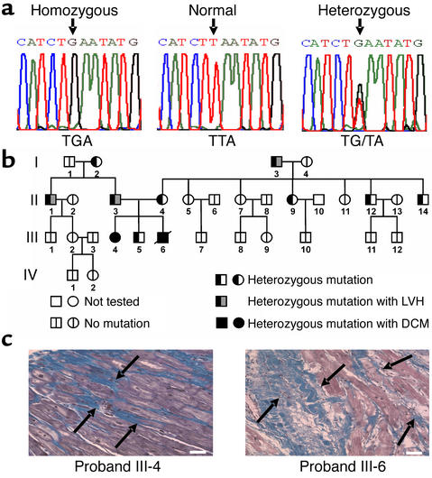
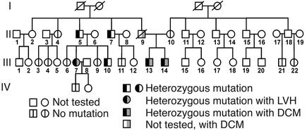
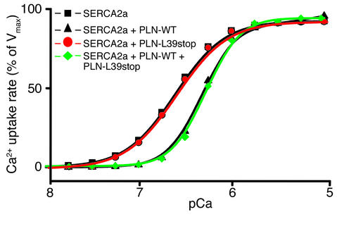
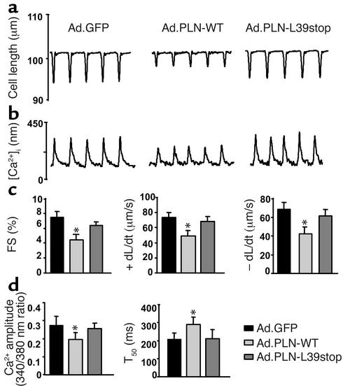
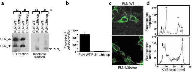
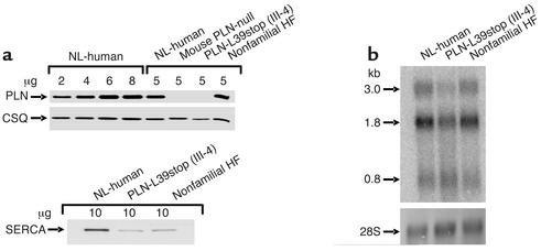
In human disease and experimental animal models, depressed Ca(2+) handling in failing cardiomyocytes is widely attributed to impaired sarcoplasmic reticulum (SR) function. In mice, disruption of the PLN gene encoding phospholamban (PLN) or expression of dominant-negative PLN mutants enhances SR and cardiac function, but effects of PLN mutations in humans are unknown. Here, a T116G point mutation, substituting a termination codon for Leu-39 (L39stop), was identified in two families with hereditary heart failure. The heterozygous individuals exhibited hypertrophy without diminished contractile performance. Strikingly, both individuals homozygous for L39stop developed dilated cardiomyopathy and heart failure, requiring cardiac transplantation at ages 16 and 27. An over 50% reduction in PLN mRNA and no detectable PLN protein were noted in one explanted heart. The expression of recombinant PLN-L39stop in human embryonic kidney (HEK) 293 cells and adult rat cardiomyocytes showed no PLN inhibition of SR Ca(2+)-ATPase and the virtual absence of stable PLN expression; where PLN was expressed, it was misrouted to the cytosol or plasma membrane. These findings describe a naturally-occurring loss-of-function human PLN mutation (PLN null). In contrast to reported benefits of PLN ablation in mouse heart failure, humans lacking PLN develop lethal dilated cardiomyopathy.


Figures

Figure 1
Figure 1
Mutation in the PLN gene and analysis of inheritance in kindred I. (a) Partial nucleotide sequences of the PLN coding region in normal subjects and in patients with dilated cardiomyopathy who were homozygous or heterozygous for the T116G transversion, which converts the codon for Leu-39 (TTA) to a stop codon (TGA). (b) Pedigree for the presence or absence of the T116G mutation in kindred I. Probands III-4 and III-6, who were homozygous for the L39stop mutation, were diagnosed with dilated cardiomyopathy and required cardiac transplantation. Squares represent males and circles represent females. A line denotes that the patient is deceased. (c) Histological analysis of explanted hearts from probands III-4 and III-6 (who were homozygous for the L39stop mutation), which were stained with Masson’s trichrome, illustrated the massive interstitial fibrosis and myocardial disarrangement (arrows). Scale bar, 50 μm.
Figure 2
Figure 2
Analysis of inheritance of the T116G mutation in kindred II. Pedigree for the presence or absence of the T116G mutation in four generations. Two brothers (probands III-13 and III-14) who were heterozygous for the L39stop mutation presented with a dilated cardiomyopathy. Their deceased father, II-9, was also diagnosed with dilated cardiomyopathy. Squares represent males and circles represent females. A line denotes that the patient is deceased.
Figure 3
Figure 3
Effect of wild-type and homozygous or heterozygous mutant PLN-L39stop on the Ca2+ affinity of SERCA2a. HEK-293 cells were cotransfected with wild-type, homozygous, or heterozygous mutant PLN cDNA and SERCA2a cDNA, and the rates of Ca2+ uptake were measured. The results are representative of two nearly identical and independent experiments. Vmax, maximum velocity of Ca2+ uptake.
Figure 4
Figure 4
Effect of PLN-L39stop mutant on adult rat cardiac myocyte mechanics and Ca2+ kinetics. Shown are representative recordings of cell shortening (a) and the Ca2+ transient (b) in cardiomyocytes isolated from adult rat hearts infected with Ad.GFP, Ad.PLN-WT, and Ad.PLN-L39stop. Myocytes were stimulated at 0.5 Hz at 25°C. (c) Percent myocyte fractional shortening (FS%), rate of contraction (+dL/dt), and rate of relaxation (–dL/dt). (d) Ca2+ transient amplitude (340/380 nm ratio) and time to 50% decay of the Ca2+ signal (T50). A minimum of 16–20 cells was studied from each of three individual preparations. Values are means ± SEM; *P < 0.05.
Figure 5
Figure 5
Expression and localization of PLN-WT and PLN-L39stop mutant in HEK-293 cells. (a) Immunoblot analyses of endoplasmic reticulum microsomes (left panel) and insoluble fractions (right panel) obtained at 24 and 48 hours after transfections. Note the absence of PLN-L39stop in the endoplasmic reticulum fraction, although detectable protein levels are found in the insoluble fraction. PLNp, PLN pentamer; PLNm, PLN monomer. (b) Quantitation of the number of fluorescent cells on PLN-WT and PLN-L39stop transfected coverslips. Cultures were transfected with equal amounts of plasmid DNA, and 48 hours later the number of fluorescent cells was counted on the entire coverslip. (c) Immunofluorescence of PLN-WT and PLN-L39stop transfected cells analyzed by confocal microscopy 48 hours after transfection. In PLN-WT transfected cells, immunofluorescence is exclusively in the endoplasmic reticulum, whereas a clearly distinct and plasma membrane–associated staining pattern is observed in the PLN-L39stop transfected cells. Scale bar, 30 μm. (d) Fluorescence intensity was quantitated using available Leica software. For these assays, a straight profile line was drawn across the center of the cell and fluorescence amplitude was plotted. Arrows mark the edge of the cell and asterisks mark the ER. In PLN-WT transfected cells (upper panel), immunofluorescence is found within the interior of the cell, whereas in PLN-L39stop transfected cells (lower panel), staining is found enriched at the outer edge of the cell.
Figure 6
Figure 6
SR proteins and PLN expression levels in explanted heart tissue of proband III-4 in kindred I. (a) Quantitative immunoblotting of PLN, SERCA2a, and calsequestrin in explanted heart tissue of homozygous proband III-4, a nonfamilial heart failure patient, and a normal donor subject as well as the PLN-null mouse. Proteins were visualized with specific antibodies against PLN, SERCA2, and calsequestrin and quantified against a standard obtained from normal human donor heart. (b) Autoradiographic images of oligonucleotide-probed Northern blot of PLN (upper panel), using cardiac RNA from normal individuals and those with PLN-L39stop (III-4) and nonfamilial heart failure. The radiolabeled 60-base oligonucleotide, antisense to the PLN coding region, recognizes the three major PLN mRNAs (0.8, 1.8, and 3.0 kb). These blots were stained with methylene blue to demonstrate the RNA integrity and loading (lower panel). CSQ, calsequestrin; HF, heart failure patient; NL, normal donor.

Comment in

References

    1. Cohn JN. Current concepts in the treatment of congestive heart failure. Cardiology. 1997;88(Suppl. 2):2–6. - PubMed
    1. Ho KK, Pinsky JL, Kannel WB, Levy D. The epidemiology of heart failure: the Framingham Study. J. Am. Coll. Cardiol. 1993;22:6A–13A. - PubMed
    1. Hasenfuss G. Alterations of calcium-regulatory proteins in heart failure. Cardiovasc. Res. 1998;37:279–289. - PubMed
    1. Beuckelmann DJ, Nabauer M, Erdmann E. Intracellular calcium handling in isolated ventricular myocytes from patients with terminal heart failure. Circulation. 1992;85:1046–1055. - PubMed
    1. Endo M. Calcium release from the sarcoplasmic reticulum. Physiol. Rev. 1977;57:71–108. - PubMed

Publication types