. 2003 Mar;111(6):877-85.
doi: 10.1172/JCI15645.

DNA damage is a novel response to sublytic complement C5b-9-induced injury in podocytes

Affiliations

DNA damage is a novel response to sublytic complement C5b-9-induced injury in podocytes

Jeffrey W Pippin et al. J Clin Invest. 2003 Mar.

Abstract

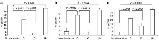
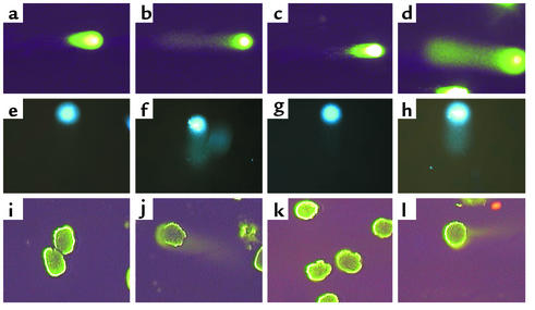
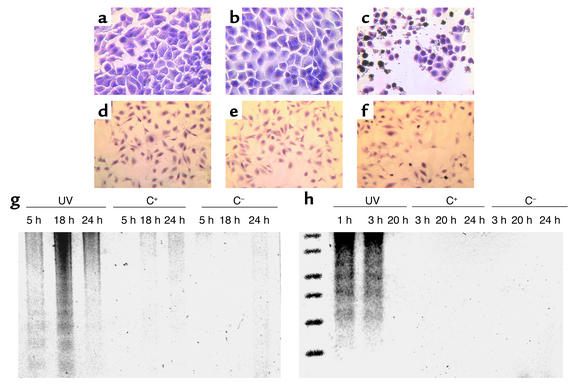
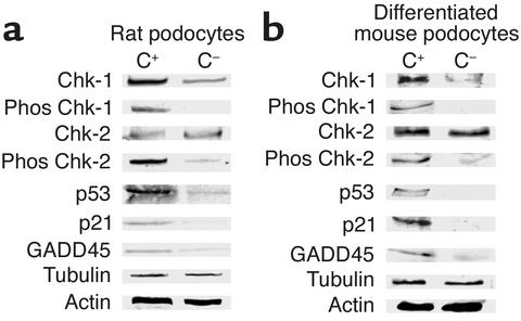
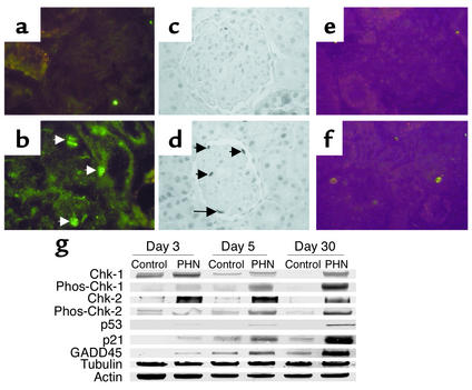
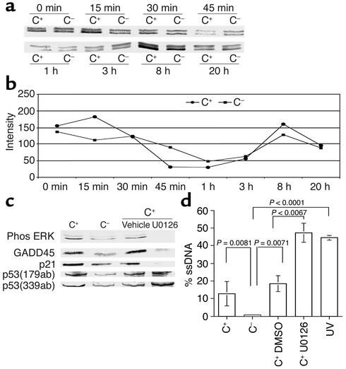
In response to Ab-complement-mediated injury, podocytes can undergo lysis, apoptosis, or, when exposed to sublytic (<5% lysis) amounts of C5b-9, become activated. Following the insertion of sublytic quantities of C5b-9, there is an increase in signaling pathways and growth factor synthesis and release of proteases, oxidants, and other molecules. Despite an increase in DNA synthesis, however, sublytic C5b-9 is associated with a delay in G(2)/M phase progression in podocytes. Here we induced sublytic C5b-9 injury in vitro by exposing cultured rat podocytes or differentiated postmitotic mouse podocytes to Ab and a complement source; we also studied the passive Heymann nephritis model of experimental membranous nephropathy in rats. A major finding was that sublytic C5b-9-induced injury caused an increase in DNA damage in podocytes both in vitro and in vivo. This was associated with an increase in protein levels for p53, the CDK inhibitor p21, growth-arrest DNA damage-45 (GADD45), and the checkpoint kinases-1 and -2. Sublytic C5b-9 increased extracellular signal-regulated kinase-1 and -2 (ERK-1 and -2), and inhibiting ERK-1 and -2 reduced the increase in p21 and GADD45 and augmented the DNA damage response to sublytic C5b-9-induced injury. These results show that sublytic C5b-9 induces DNA damage in vitro and in vivo and may explain why podocyte proliferation is limited following immune-mediated injury.


Figures

Figure 1
Figure 1
Sublytic C5b-9 causes DNA damage in podocytes in vitro. ssDNA damage was measured by the DNA-precipitation assay in proliferating rat (a), quiescent rat (b), and differentiated postmitotic mouse (c) podocytes. DNA damage was barely detected in control podocytes not exposed to Ab (lane 1) and in control cells exposed to Ab without a complement source (C, lane 3). In contrast, DNA damage was significantly increased in podocytes following sublytic C5b-9 injury when exposed to Ab and a complement source (C+, lane 2). DNA damage was detected in positive control cells exposed to UV irradiation (UV, lane 4). These results show that sublytic C5b-9 injury causes DNA damage in cultured rat and mouse podocytes.
Figure 2
Figure 2
Comet assay. DNA damage was measured in rat (ad) and mouse (eh) podocytes in vitro using the comet assay. (a and e) DNA damage was not detected in normal podocytes not exposed to Ab or a complement source. (b and f) In podocytes exposed to Ab and complement, the characteristic “tail” of damaged DNA is seen migrating away from the nucleus. (c and g) In contrast, DNA damage was not detected in control cells exposed to Ab without a complement source. (d and h) In UV-irradiated cells, used as a positive control, DNA damage was detected. The comet assay was also performed on glomeruli isolated from control and PHN rats. (il) DNA damage was not detected in control rats injected with normal sheep serum (i) nor in PVG C rats injected with anti-Fx1A Ab (k). DNA damage was detected in Sprague-Dawley PHN (j) and in PVG C+ (l) rats injected with anti-Fx1A Ab.
Figure 3
Figure 3
Sublytic C5b-9 does not induce apoptosis in podocytes. af show TUNEL staining, a marker of apoptosis. Apoptosis was not detected in control rat (a) and mouse (d) podocytes exposed to Ab without a complement source, nor in experimental rat (b) and mouse (e) cells exposed to Ab with a complement source. In contrast, TUNEL staining is increased in rat (c) and mouse (f) podocytes exposed to UV irradiation used as a positive control. The suicide track DNA ladder assay is shown in rat (g) and mouse (h) podocytes. In contrast to DNA fragmentation in podocytes exposed to UV irradiation (lanes 1, 2, 3), DNA fragmentation was not detected in cells exposed to Ab with (C+) (lanes 4, 5, 6) and without (C) (lanes 7, 8, 9) a complement source.
Figure 4
Figure 4
Sublytic C5b-9 increases CHK-1, CHK-2, p53, p21, and GADD45 expression in podocytes in vitro. Western blot analysis was used to measure proteins from in vitro rat (a) and mouse (b) podocytes exposed to Ab with a complement source (C+) and without a complement source (C). C5b-9 increased the protein levels of total CHK-1, phosphorylated (phos) CHK-1, phosphorylated CHK-2, p53, p21, and GADD45. The housekeeping proteins, actin and tubulin, showed that protein loading was equivalent.
Figure 5
Figure 5
DNA damage proteins increase in PHN rats. Immunostaining for p53 (a), p21 (c), and phosphorylated CHK-2 (e) was not detected in control rats injected with normal sheep serum. In contrast, p53 (b), p21 (d), and phosphorylated CHK-2 immunostaining (f) increased in Sprague-Dawley rats with PHN (arrows indicate positive-stained podocytes). g The results of Western blot analysis performed on isolated glomerular protein from control and PHN rats. In PHN animals, the levels for phosphorylated (phos) CHK-1 and CHK-2, p53, p21, and GADD45 increased compared with controls. The housekeeping proteins, actin and tubulin, were used to ensure equal protein loading.
Figure 6
Figure 6
ERK regulates p21 and GADD45 following sublytic C5b-9 attack. (a) Phosphorylated ERK was measured by Western blot analysis in control (C) and C5b-9–attacked (C+) podocytes, and (b) densitometry was used to quantitate the changes compared with total ERK levels. Phosphorylated ERK increased within 15 min of sublytic C5b-9 attack, and this normalized by 1 h. This was followed by a second increase in phosphorylated ERK at 8 h. (c) Inhibiting phospho-ERK with U0126 reduced GADD45 and p21. (d) Inhibiting ERK-1 and -2 with U0126 augments the DNA damage induced by sublytic C5b-9 (lane 4) compared with controls (lane 3).

References

    1. Couser WG. Mediation of immune glomerular injury. J. Am. Soc. Nephrol. 1990;1:13–29. - PubMed
    1. Cybulsky AV, Monge JC, Papillon J, McTavish AJ. Complement C5b-9 activates cytosolic phospholipase A2 in glomerular epithelial cells. Am. J. Physiol. 1995;269:F739–F749. - PubMed
    1. Torbohm I, et al. C5b-8 and C5b-9 modulate the collagen release of human glomerular epithelial cells. Kidney Int. 1990;37:1098–1104. - PubMed
    1. Hansch GM, Seitz M, Betz M. Effect of the late complement components C5b-9 on human monocytes: release of prostanoids, oxygen radicals and of a factor inducing cell proliferation. Int. Arch. Allergy Appl. Immunol. 1987;82:317–320. - PubMed
    1. Lovett DH, Haensch G-M, Goppelt M, Resch KDG. Activation of glomerular mesangial cells by the terminal membrane attack complex of complement. J. Immunol. 1987;138:2413–2480. - PubMed

Publication types

MeSH terms