. 2003 Apr;23(7):2264-76.
doi: 10.1128/MCB.23.7.2264-2276.2003.

The Delta Np63 alpha phosphoprotein binds the p21 and 14-3-3 sigma promoters in vivo and has transcriptional repressor activity that is reduced by Hay-Wells syndrome-derived mutations

Affiliations

The Delta Np63 alpha phosphoprotein binds the p21 and 14-3-3 sigma promoters in vivo and has transcriptional repressor activity that is reduced by Hay-Wells syndrome-derived mutations

Matthew D Westfall et al. Mol Cell Biol. 2003 Apr.

Abstract

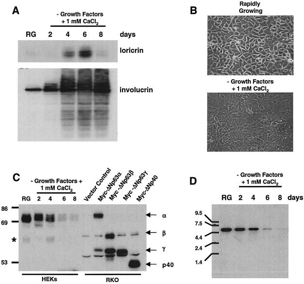
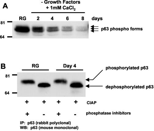
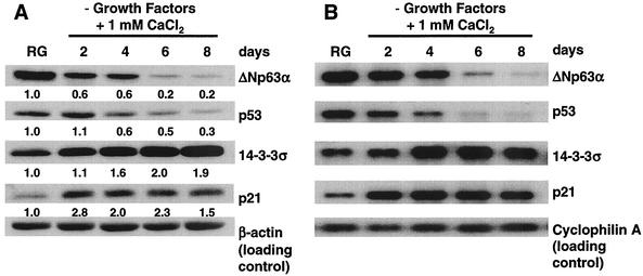
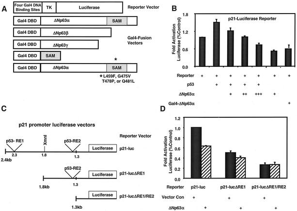
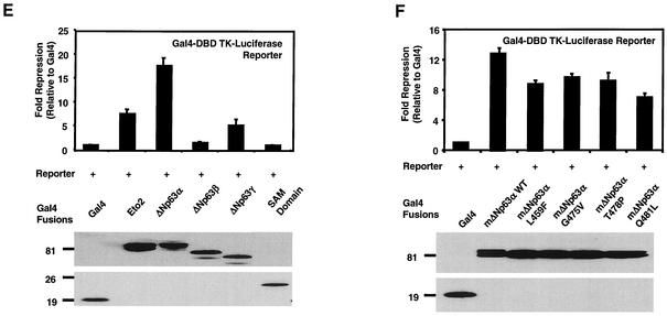
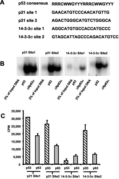
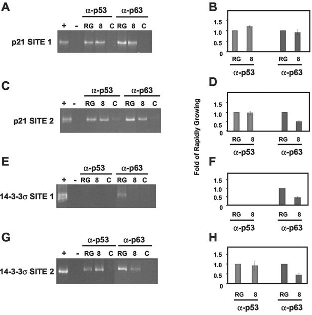
p63 is a recently identified homolog of p53 that is found in the basal layer of several stratified epithelial tissues such as the epidermis, oral mucosa, prostate, and urogenital tract. Studies with p63(-/-) mice and analysis of several human autosomal-dominant disorders with germ line p63 mutations suggest p63 involvement in maintaining epidermal stem cell populations. The p63 gene encodes six splice variants with reported transactivating or dominant-negative activities. The goals of the current study were to determine the splice variants that are expressed in primary human epidermal keratinocytes (HEKs) and the biochemical activity p63 has in these epithelial cell populations. We found that the predominant splice variant expressed in HEKs was Delta Np63 alpha, and it was present as a phosphorylated protein. During HEK differentiation, Delta Np63 alpha and p53 levels decreased, while expression of p53 target genes p21 and 14-3-3 sigma increased. Delta Np63 alpha had transcriptional repressor activity in vitro, and this activity was reduced in Delta Np63 alpha proteins containing point mutations, corresponding to those found in patients with Hay-Wells syndrome. Further, we show that Delta Np63 alpha and p53 can bind the p21 and 14-3-3 sigma promoters in vitro and in vivo, with decreased binding of p63 to these promoters during HEK differentiation. These data suggest that Delta Np63 alpha acts as a transcriptional repressor at select growth regulatory gene promoters in HEKs, and this repression likely plays an important role in the proliferative capacity of basal keratinocytes.


Figures

FIG. 1.
FIG. 1.
Differentiation-induced modulation of ΔNp63α protein levels in primary HEKs. HEKs were induced to differentiate as described in Materials and Methods. (A) Western analysis of HEK lysates from rapidly growing and differentiating cells for expression of the terminal differentiation markers loricrin and involucrin. (B) Micrograph of rapidly growing HEKs and HEKs at day 4 after induction of differentiation. (C) Western analysis with antibody Ab-1 for p63 expression in rapidly growing and differentiating HEKs. Myc epitope-tagged ΔNp63 splice variants were ectopically expressed in RKO cells, and lysates were analyzed to serve as molecular weight markers for comparison to HEK p63. The asterisk to the left of the blot aligns with a cross-reactive band that may represent ΔNp63β expression. (D) Northern analysis of p63 transcript expression in HEKs differentiated as in panel A. The results shown are representative of three independent experiments with separate primary cultures of HEKs.
FIG. 2.
FIG. 2.
ΔNp63α is a phosphoprotein. (A) Protein lysates were prepared from rapidly growing and differentiating HEKs and analyzed by Western blotting with mouse monoclonal antibody Ab-1. Arrows denote three differentially migrating ΔNp63α forms. (B) p63 was immunoprecipitated with rabbit polyclonal antibody H129, and immoprecipitates were treated with CIAP in the presence or absence of phosphatase inhibitors. Immunoprecipitated protein was analyzed by Western blotting for p63 by using mouse monoclonal antibody Ab-1. Resolution of the multiple bands by SDS-PAGE in Fig. 2B was not possible due to the percentage of the acrylamide used. The results shown are representative of four independent experiments.
FIG. 3.
FIG. 3.
Differentiation-induced changes in select protein and mRNA levels in primary HEKs. HEKs were induced to differentiate as described in Materials and Methods. (A) Western analysis of p63 (Ab-1), p53 (Ab-2), 14-3-3σ (N-14), and p21 (Ab-1) in rapidly growing and differentiating HEKs with the antibodies listed in Materials and Methods. Note that p63 migrates as a single band due to the use of electrophoresis conditions that allow for analysis of the molecular weight range of proteins shown in panel A. The numbers below the Western panels represent the fold change relative to rapidly growing HEKs. (B) Northern analysis of transcripts for proteins shown in panel A. The results are representative of three independent experiments with independent primary cultures of HEKs.
FIG. 4.
FIG. 4.
ΔNp63α represses transcription. (A) Schematic representing vectors used in panels B, D, E, and F. Abbreviations: DBD, DNA-binding domain; TK, thymidine kinase. The SAM mutants were L459F, G475V, T478P, and Q481L. (B) Analysis of ΔNp63α and Gal4-ΔNp63α activity in a p21 promoter-luciferase reporter assay. HCT116 cells were transfected with a p21 promoter-luciferase reporter construct (containing 2,400 bp of p21 promoter sequence) and expression vector encoding either p53, ΔNp63α, or Gal4-ΔNp63α. +, ++, and +++, indicate 1:1, 1:3, and 1:10 ratios of p53 to ΔNp63α expression vectors, respectively. (C) Schematic representing reporter vectors used in panel D. Abbreviations: p53-RE, p53-response element; luc, luciferase. (D) HCT116 cells were transfected as in panel B with the indicated reporter vectors and an empty expression vector (Vector Con) or one expressing ΔNp63α. All values were normalized to those generated with lysates prepared from cells cotransfected with the full-length p21-luciferase reporter vector and the empty expression vector, pCEP4. (E) 293 cells were transfected with the luciferase reporter vector in panel A and the indicated Gal4 fusion vectors. Gal4 alone served as the negative control, and Gal4-ETO2 served as the positive control. (F) 293 cells were transfected with expression vectors encoding murine Gal4-ΔNp63α and Gal4-ΔNp63α proteins containing the indicated SAM domain point mutations. All luciferase assays were normalized for transfection efficiency with a renilla reporter vector. Western analyses with a Gal4-specific antibody were performed to verify protein expression, and results are shown in the lower portions of panels E and F. The results shown are representative of five independent experiments performed each time in triplicate, and error bars indicate the standard deviation.
FIG. 4.
FIG. 4.
ΔNp63α represses transcription. (A) Schematic representing vectors used in panels B, D, E, and F. Abbreviations: DBD, DNA-binding domain; TK, thymidine kinase. The SAM mutants were L459F, G475V, T478P, and Q481L. (B) Analysis of ΔNp63α and Gal4-ΔNp63α activity in a p21 promoter-luciferase reporter assay. HCT116 cells were transfected with a p21 promoter-luciferase reporter construct (containing 2,400 bp of p21 promoter sequence) and expression vector encoding either p53, ΔNp63α, or Gal4-ΔNp63α. +, ++, and +++, indicate 1:1, 1:3, and 1:10 ratios of p53 to ΔNp63α expression vectors, respectively. (C) Schematic representing reporter vectors used in panel D. Abbreviations: p53-RE, p53-response element; luc, luciferase. (D) HCT116 cells were transfected as in panel B with the indicated reporter vectors and an empty expression vector (Vector Con) or one expressing ΔNp63α. All values were normalized to those generated with lysates prepared from cells cotransfected with the full-length p21-luciferase reporter vector and the empty expression vector, pCEP4. (E) 293 cells were transfected with the luciferase reporter vector in panel A and the indicated Gal4 fusion vectors. Gal4 alone served as the negative control, and Gal4-ETO2 served as the positive control. (F) 293 cells were transfected with expression vectors encoding murine Gal4-ΔNp63α and Gal4-ΔNp63α proteins containing the indicated SAM domain point mutations. All luciferase assays were normalized for transfection efficiency with a renilla reporter vector. Western analyses with a Gal4-specific antibody were performed to verify protein expression, and results are shown in the lower portions of panels E and F. The results shown are representative of five independent experiments performed each time in triplicate, and error bars indicate the standard deviation.
FIG. 5.
FIG. 5.
Relative binding affinities of p53 and ΔNp63α for p53 consensus sites in the p21 and 14-3-3σ promoters. (A) The p53 consensus binding sequence and p53 binding sites in the p21 and 14-3-3σ promoters. Abbreviations: R, purine; Y, pyrimidine; W, adenine or thymine. (B) H1299 cells were transfected with myc-tagged p53 or ΔNp63α, and protein lysates were quantified by using the Fluor-S Max MultiImager. Based on Fluor-S Max quantification, equal amounts of myc-tagged p53 and myc-tagged ΔNp63α were immunoprecipitated with a myc epitope antibody. Immunoprecipitated p53 and ΔNp63α were assayed for their ability to bind radiolabeled oligonucleotides representing p53-binding sites in the p21 and 14-3-3σ promoters as described in Materials and Methods. (C) Bound oligonucleotides were separated on acrylamide gels, exposed for autoradiography, and quantified. Each autoradiograph shows one representative result of at least three independent experiments that are quantified and displayed with the standard deviation.
FIG. 6.
FIG. 6.
ChIP analyses for p53 and p63 binding at the 14-3-3σ and p21 promoters during keratinocyte differentiation. Abbreviations: RG, rapidly growing; 8, day 8 after induction of differentiation; C, control. HEKs were induced to differentiate as described in Materials and Methods. At the time of harvest, HEKs were treated with formaldehyde (X-Link) and processed as described in Materials and Methods. The DNA immunoprecipitated with p53- or p63-specific antibodies was PCR amplified by using primers flanking the p53 binding sites in the p21 (A and C) and 14-3-3σ (E and G) promoters. DNA fragments generated by PCR were resolved by PAGE, the gels were stained with ethidium bromide, and the PCR products were quantified by densitometry. Lanes marked “+” indicate PCR products that were generated by using DNA template derived from total genomic DNA harvested from rapidly growing HEKs. Lanes marked “−” indicate the absence of DNA input for the PCR. The lanes marked “C” indicate PCRs performed with templates immunoprecipitated with antibodies specific to cyclin B1 and Bax for p53 and p63 analyses, respectively. Each ethidium bromide-stained gel shows one representative result of at least three independent experiments that are quantified and displayed with standard deviations in the corresponding panels B, D, F, and H.

References

    1. Amann, J. M., J. Nip, D. K. Strom, B. Lutterbach, H. Harada, N. Lenny, J. R. Downing, S. Meyers, and S. W. Hiebert. 2001. ETO, a target of t(8;21) in acute leukemia, makes distinct contacts with multiple histone deacetylases and binds mSin3A through its oligomerization domain. Mol. Cell. Biol. 21:6470-6483. - PMC - PubMed
    1. Augustin, M., C. Bamberger, D. Paul, and H. Schmale. 1998. Cloning and chromosomal mapping of the human p53-related KET gene to chromosome 3q27 and its murine homolog Ket to mouse chromosome 16. Mamm. Genome 9:899-902. - PubMed
    1. Barrandon, Y., and H. Green. 1985. Cell size as a determinant of the clone-forming ability of human keratinocytes. Proc. Natl. Acad Sci. USA 82:5390-5394. - PMC - PubMed
    1. Barrandon, Y., and H. Green. 1987. Three clonal types of keratinocyte with different capacities for multiplication. Proc. Natl. Acad. Sci. USA 84:2302-2306. - PMC - PubMed
    1. Celli, J., P. Duijf, B. Hamel, M. Bamshad, B. Kramer, A. Smits, R. Newbury-Ecob, R. Hennekam, G. Van Buggenhout, A. van Haeringen, C. Woods, A. van Essen, R. de Waal, G. Vriend, D. Haber, A. Yang, F. McKeon, H. Brunner, and H. van Bokhoven. 1999. Heterozygous germline muations in the p53 homolog p63 are the cause of EEC syndrome. Cell 99:143-153. - PubMed

Publication types

MeSH terms