. 2003 Apr;23(7):2407-14.
doi: 10.1128/MCB.23.7.2407-2414.2003.

Caveolin-induced activation of the phosphatidylinositol 3-kinase/Akt pathway increases arsenite cytotoxicity

Affiliations

Caveolin-induced activation of the phosphatidylinositol 3-kinase/Akt pathway increases arsenite cytotoxicity

Sonsoles Shack et al. Mol Cell Biol. 2003 Apr.

Abstract

The inhibitory effect of caveolin on the cellular response to growth factor stimulation is well established. Given the significant overlap in signaling pathways involved in regulating cell proliferation and stress responsiveness, we hypothesized that caveolin would also affect a cell's ability to respond to environmental stress. Here we investigated the ability of caveolin-1 to modulate the cellular response to sodium arsenite and thereby alter survival of the human cell lines 293 and HeLa. Cells stably transfected with caveolin-1 were found to be much more sensitive to the toxic effects of sodium arsenite than either untransfected parental cells or parental cells transfected with an empty vector. Unexpectedly, the caveolin-overexpressing cells also exhibited a significant activation of the phosphatidylinositol 3-kinase (PI3K)/Akt pathway, which additional studies suggested was likely due to decreased neutral sphingomyelinase activity and ceramide synthesis. In contrast to its extensively documented antiapoptotic influence, the elevated activity of Akt appears to be important in sensitizing caveolin-expressing cells to arsenite-induced toxicity, as both pretreatment of cells with the PI3K inhibitor wortmannin and overexpression of a dominant-negative Akt mutant markedly improved the survival of arsenite-treated cells. This death-promoting influence of the PI3K/Akt pathway in caveolin-overexpressing cells appeared not to be unique to sodium arsenite, as wortmannin pretreatment also resulted in increased survival in the presence of H(2)O(2). In summary, our results indicate that caveolin-induced upregulation of the PI3K/Akt signaling pathway, which appears to be a death signal in the presence of arsenite and H(2)O(2), sensitizes cells to environmental stress.


Figures

FIG. 1.
FIG. 1.
Caveolin overexpression in 293 cells leads to elevation in phospho-Akt. 293 Cells stably transfected to overexpress caveolin-1 were clonally selected by using G418, and phospho(p)-Akt levels were assessed by Western blotting. Shown are nine clonal isolates along with pooled 293 cells transfected with an insertless vector (pcDNA3.1). Fifty-microgram protein aliquots were resolved in 10% polyacrylamide SDS gels, transferred onto polyvinylidene difluoride membranes, and hybridized by using an anti-caveolin-1 antibody that recognizes both the monomeric and oligomeric forms of caveolin-1. Membranes were stripped and hybridized with an antibody recognizing phosphorylated Akt (p-Akt), and actin is shown as an internal control.
FIG. 2.
FIG. 2.
Inhibition of ceramide synthesis by caveolin likely contributes to elevating Akt phosphorylation. (A) Time-dependent reduction of Akt phosphorylation in 293-Cav25 cells treated with 40 μM C2 ceramide. (B) Dose-dependent reduction of Akt phosphorylation in 293-Cav25 cells treated for 2 h with the indicated doses of C2 ceramide. Levels of Akt protein are also shown as an internal control. (C) nSMase (SM) enzymatic activity in 293-V and 293-Cav25 cells was assessed by monitoring cleavage of radiolabeled sphingomyelin, the precursor of ceramide, as described in Materials and Methods.
FIG. 3.
FIG. 3.
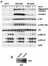
Caveolin-induced changes in Akt and MAP kinase signaling pathways after exposure of 293 cells to arsenite. (A) 293-V, 293-Cav25, and 293-Cav31 cells were treated with 25 μM arsenite for the indicated times, whereupon Western blot analysis was carried out to assess the levels of oligomerized caveolin, monomeric caveolin, p-Akt, Akt, p-GSK-3 α/β, p-ERK, p-JNK, and p-p38 in 50 μg of whole-cell lysate. (B) 293-V and 293-Cav25 cells were harvested, and Akt was immunoprecipitated from lysates with a polyclonal anti-Akt antibody. Akt kinase activity was assessed by using histone H2B as a substrate.
FIG. 4.
FIG. 4.
Elevated Akt phosphorylation in caveolin-overexpressing cells is likely due to elevated PI3K activity and not to diminished PTEN expression. (A) Western blot analysis of p-Akt expression in 293-V and 293-Cav25 cells following either no treatment or treatment for 1 h with an inhibitor of growth factor receptors (20 μM AG1296, 1 μM AG1478, or 300 μM suramin), the MEK1/2 inhibitor U0126 (20 μM), or the PI3K inhibitor LY294002 (25 μM). (B) Western blot analysis of PTEN levels in 293-V, 293-Cav25, and 293-Cav31 cells treated with 25 μM arsenite for the times indicated.
FIG. 5.
FIG. 5.
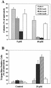
Decreased survival of caveolin-overexpressing 293 cells following arsenite treatment. (A) Clonogenicity assay to monitor the sensitivity of 293-V, 293-Cav25, and 293-Cav31 cells to 5 and 10 μM arsenite. Cells were treated with arsenite for 24 h, and surviving colonies were stained and scored as described in Materials and Methods. Approximately 1,000 cells were evaluated for each data point. (B) Trypan blue exclusion assay to monitor survival of 293 cells expressing different levels of caveolin following treatment with 25 μM arsenite for 24 h. Data shown in both panels are the means and standard deviations from three independent experiments.
FIG. 6.
FIG. 6.
Differential sensitivity of arsenite-treated HeLa cells expressing different levels of caveolin. (A) HeLa cells transfected with either empty vector pcDNA3.1 (HeLa-V) or pcDNA3.1-caveolin-1 (HeLa-C1) to overexpress caveolin-1 were treated with arsenite, and caveolin-1 and p-Akt expression were subsequently evaluated by Western blot analysis. (B) HeLa-V and HeLa-Cav1 cells were harvested, and Akt was immunoprecipitated. Akt kinase activity was evaluated by using histone H2B as a substrate. (C) Graph depicting the quantitation of clonogenicity assays following exposure to the doses of arsenite shown for 24 h. (D) Photograph illustrating the different numbers of colonies formed by HeLa-V and HeLa-C1 populations after treatment with 10 μM arsenite for 24 h, replating for clonogenicity assay, and staining with crystal violet. Data represent the means and standard deviations from three independent experiments.
FIG. 7.
FIG. 7.
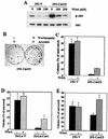
Effect of wortmannin on the sensitivity of caveolin-overexpressing cells to arsenite and H2O2. (A) Western blotting showing the inhibition of Akt phosphorylation by 100 nM wortmannin (Wort) treatment for 1 h. Total Akt levels are also shown for comparison. (B) Photograph illustrating the different numbers of colonies formed by 293-C25 populations after treatment with 10 μM arsenite for 24 h in the absence or presence of wortmannin. Graphs depict the quantitation of clonogenicity assays when cells are stressed in the absence or presence of wortmannin (100 nM) for 293-V and 293-Cav25 cells exposed to 10 μM arsenite (C); HeLa-V and HeLa-C1 cells treated with 10 μM arsenite (D); 293-V and 293-Cav25 exposed to 10 μM H2O2 (E). Clonogenicity assays were carried out as described in Materials and Methods. An asterisk indicates that the result is not statistically significant by t test analysis (P > 0.05). Data represent the means and standard deviations from three independent experiments.
FIG. 8.
FIG. 8.
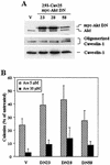
Influence of dominant-negative Akt on the survival of 293-Cav25 cells. (A) Western blot analysis of the expression of myc-Akt DN in selected clones (Cav25-DN23, Cav25-DN28, and Cav25-DN58) obtained from expressing pUSE-Akt1 (K179M mutant) or the pUSE empty vector (Cav25-V). Anti-Akt antibodies allowed for the detection of both endogenous Akt and myc-Akt DN expression (the latter exhibiting higher molecular weight than that of endogenous Akt). In addition, oligomerized and monomeric forms of caveolin-1 are also shown. (B) Graph depicting the number of colonies formed by the Cav25 Akt DN clones selected and by a cellular pool expressing the pUSE vector after treatment with 5 and 10 μM arsenite (Ars) for 24 h. Colonies were stained and evaluated as described in Materials and Methods. Differences in the survival of Cav25-DN23 and Cav25-DN28 with respect to Cav25-V were statistically significant by t test analysis (P < 0.05). These results represent the means and standard deviations from three independent experiments.

References

    1. Alessandrini, A., S. Namura, M. A. Molkowitz, and J. V. Bonaventre. 1999. MEK1 protein kinase inhibition protects against damage resulting from focal cerebral ischemia. Proc. Natl. Acad. Sci. USA 96:12866-12869. - PMC - PubMed
    1. Anderson, R. G. W. 1998. The caveolae membrane system. Annu. Rev. Biochem. 67:199-225. - PubMed
    1. Arcaro, A., and M. P. Wymann. 1993. Wortmannin is a potent phosphatidylinositol 3-kinase inhibitor: the role of phosphatidylinositol 3,4,5-trisphosphate in neutrophil responses. Biochem. J. 296:297-301. - PMC - PubMed
    1. Bagla, P., and J. Kaiser. 1996. India's spreading health crisis draws global arsenic experts. Science 274:174-175. - PubMed
    1. Barchowsky, A., E. J. Dudek, M. D. Treadwell, and K. E. Wetterhan. 1996. Arsenic induces oxidant stress and NF-kappa B activation in cultured aortic endothelial cells. Free Radic. Biol. Med. 21:783-790. - PubMed

MeSH terms

LinkOut - more resources