. 2003 Apr;23(7):2425-37.
doi: 10.1128/MCB.23.7.2425-2437.2003.

Myocardin is a critical serum response factor cofactor in the transcriptional program regulating smooth muscle cell differentiation

Affiliations

Myocardin is a critical serum response factor cofactor in the transcriptional program regulating smooth muscle cell differentiation

Kevin L Du et al. Mol Cell Biol. 2003 Apr.

Abstract

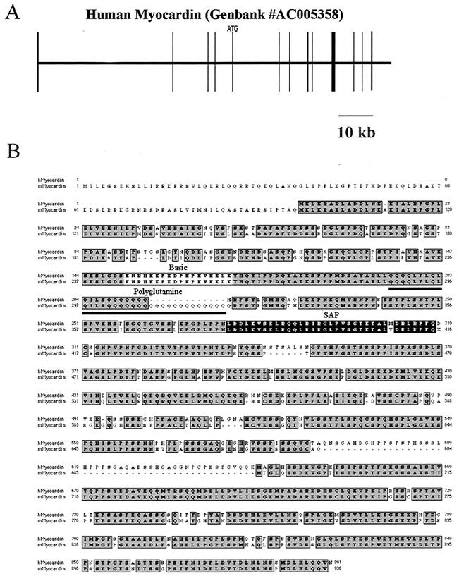
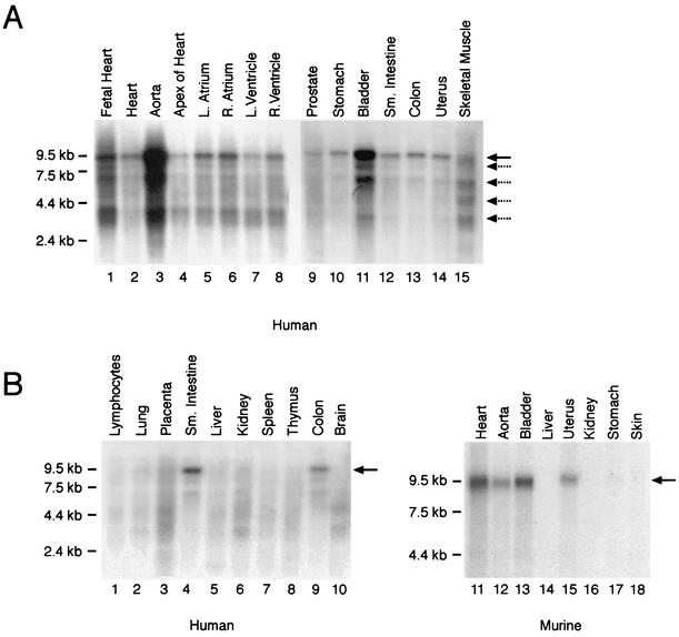
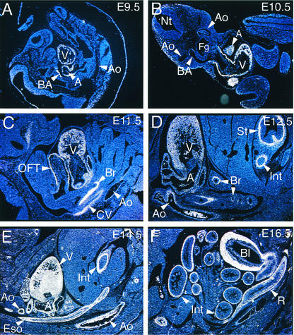
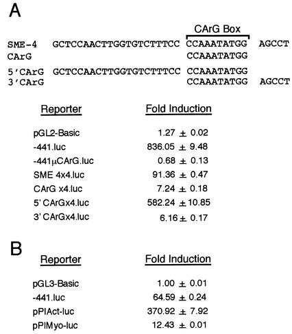
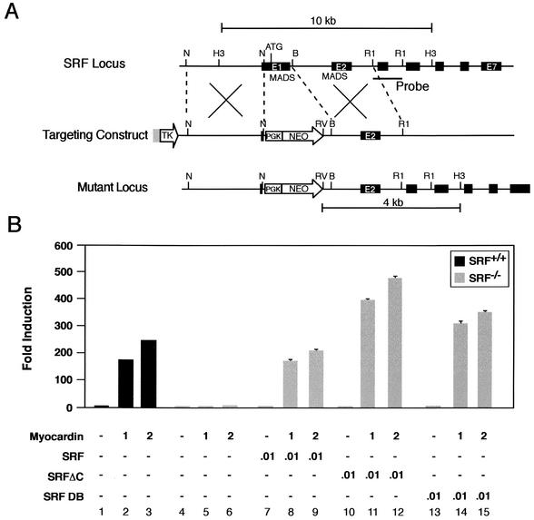
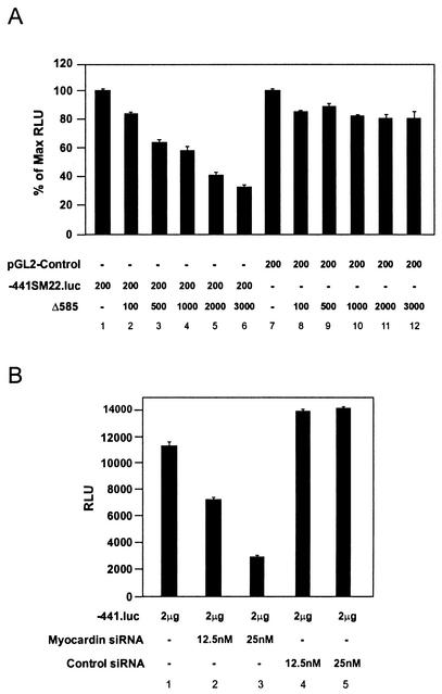
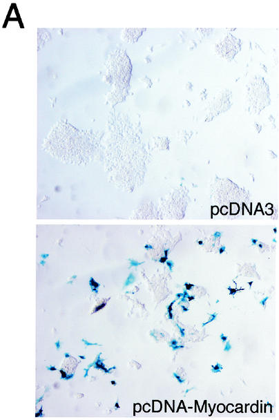
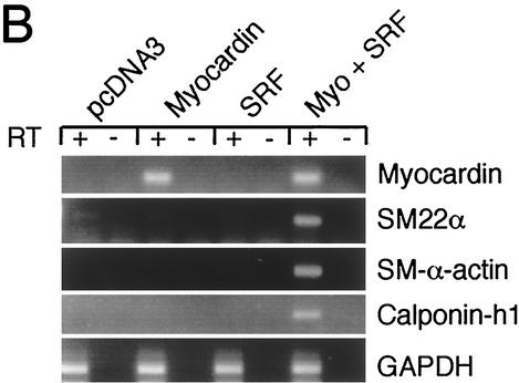
The SAP family transcription factor myocardin functionally synergizes with serum response factor (SRF) and plays an important role in cardiac development. To determine the function of myocardin in the smooth muscle cell (SMC) lineage, we mapped the pattern of myocardin gene expression and examined the molecular mechanisms underlying transcriptional activity of myocardin in SMCs and embryonic stem (ES) cells. The human and murine myocardin genes were expressed in vascular and visceral SMCs at levels equivalent to or exceeding those observed in the heart. During embryonic development, the myocardin gene was expressed abundantly in a precise, developmentally regulated pattern in SMCs. Forced expression of myocardin transactivated multiple SMC-specific transcriptional regulatory elements in non-SMCs. By contrast, myocardin-induced transactivation was not observed in SRF(-/-) ES cells but could be rescued by forced expression of SRF or the SRF DNA-binding domain. Furthermore, expression of a dominant-negative myocardin mutant protein or small-interfering-RNA-induced myocardin knockdown significantly reduced SM22 alpha promoter activity in SMCs. Most importantly, forced expression of myocardin activated expression of the SM22 alpha, smooth muscle alpha-actin, and calponin-h1 genes in undifferentiated mouse ES cells. Taken together, these data demonstrate that myocardin plays an important role in the SRF-dependent transcriptional program that regulates SMC development and differentiation.


Figures

FIG. 1.
FIG. 1.
Primary structure of the human myocardin gene and alignment of the deduced amino acid sequences of the human and murine myocardin proteins. (A) A schematic map of the human myocardin gene structure. Performing a Blast search of the GenBank database identified the human myocardin gene. The myocardin gene is located on human chromosome 17 and is identified as GenBank accession number AC005358. Exons are shown as vertical boxes. The initiation codon (ATG) in exon 5 is shown. A 10-kb scale is shown below the map. (B) Alignment of the deduced amino acid sequences of the deduced human and murine myocardin proteins. The deduced amino acid sequence of mouse myocardin is shown below the sequence of the human myocardin protein. The conserved amino acid residues are shaded dark gray. The conserved basic domain (light gray box), polyglutamine tract (underline), and SAP box (black box) are shown.
FIG. 2.
FIG. 2.
In vivo tissue distribution of myocardin gene expression. (A) Northern blot analyses of muscle cell-containing tissues. Membranes containing 2 μg of poly(A)+ RNA per lane isolated from embryonic and adult human tissues were hybridized to the radiolabeled human myocardin cDNA probe (1 week of exposure). RNA markers are shown to the left of the blot (in kilobases). The human myocardin probe hybridized to a predominant mRNA species of approximately 9.5 kb (black arrow). In addition, four to five additional low-abundance transcripts (dashed arrows) migrating between 8.5 and 3.0 kb were observed. Myocardin mRNA was observed in the heart and smooth muscle cell-containing tissues, including the aorta, stomach, bladder, small intestine, colon, and uterus. (B) Northern blot analysis of mRNA samples isolated from adult human (left panel) and murine (right panel) tissues hybridized to the radiolabeled human (left panel) and murine (right panel) myocardin cDNA probes (1 week of exposure). Myocardin mRNA (black arrow) was observed in human and murine heart and SMC-containing tissues but not in other tissues.
FIG. 3.
FIG. 3.
Myocardin gene is expressed in a developmentally regulated fashion in vascular and visceral SMCs. In situ hybridization analyses were performed on staged CD-1 mouse embryos with a 35S-labeled myocardin antisense probe. (A) In this sagittal section of an E9.5 embryo, hybridization of the myocardin riboprobe (white signal) to the ventricular chamber (V), common atria (A), and branchial arch artery (BA) was observed. Myocardin mRNA was not detectable in the dorsal aorta (Ao) at E9.5. Magnification, ×5. (B) In this transverse section of an E10.5 embryo, hybridization of the myocardin riboprobe to the ventricular chamber (V), atria (A), and cardiac outflow tract was observed. A faint signal, above background levels, was also observed in the third branchial arch artery (BA) near its communication with the aorta (Ao). Magnification, ×5. (C) In this sagittal section of the thoracic cavity of an E11.5 embryo, hybridization of the myocardin riboprobe to the ventricle (V), atria, cardiac outflow tract (OFT), primitive bronchi (Br) of the lung bud, common cardinal vein (CV), and dorsal aorta (Ao) was observed. Magnification, ×12.5. (D) In this sagittal section of the thoracic cavity and abdomen of an E12.5 embryo, hybridization of the myocardin riboprobe to the ventricle (V), atria (A), bronchi (Br) in the lung bud, aorta (Ao), stomach (St), and intestine (Int) was observed. Magnification, ×12.5. (E) In this sagittal section of the thoracic cavity and abdomen of an E14.5 embryo, hybridization of the myocardin riboprobe to the ventricle (V), atria (A), aorta (Ao), esophagus (Eso), and intestine (Int) was observed. Intense hybridization of the riboprobe to the urogenital ridge was also observed at this stage (not shown). Magnification, ×5. (F) In this sagittal section of the abdomen of an E16.5 embryo, hybridization of the myocardin riboprobe to the small and large intestine (Int), rectum (R), and muscular wall of the bladder (Bl) was observed. Magnification, ×5. No hybridization of the control sense myocardin riboprobe was observed.
FIG. 4.
FIG. 4.
Myocardin-induced transactivation of multiple SMC-specific transcriptional regulatory elements in COS-7 and undifferentiated mouse ES cells. (A) cis-acting mechanisms regulating myocardin-induced transactivation of the SM22α promoter in COS-7 cells. The nucleotide sequence of SME-4 (bp −171 to −136) and SME-4 deletion mutants CArG (bp −150 to −141), 5′CArG (bp −171 to −141), and 3′CArG (bp −151 to −136) are shown. COS-7 cells were cotransfected with the pcDNA-Myocardin expression plasmid, the phRL-TK(-Int) reference plasmid, and the indicated reporter plasmid. Luciferase activities were measured 48 h posttransfection. Myocardin-induced transcriptional activation of luciferase reporter plasmids placed under the transcriptional control of the 441-bp mouse SM22α promoter (−441.luc), the SM22α promoter containing mutations that abolish SRF binding (−441μCArG.luc), the 90-bp SM22α promoter linked to four copies of SME-4 (SME4×4.luc), the 90-bp SM22α promoter linked to four copies of the CArG oligonucleotide (CarGx4.luc), the 90-bp SM22α promoter linked to four copies of the 5′CArG oligonucleotide (5′CarGx4.luc), and the 90-bp SM22α promoter linked to four copies of the 3′CArG oligonucleotide (3′CarGx4.luc). Luciferase activity is reported as induction of luciferase activity observed when each reporter plasmid was cotransfected with pcDNA-Myocardin relative to the activity observed with pcDNA3. Results are expressed as the mean ± SEM. (B) Myocardin-induced transactivation of multiple SMC-specific transcriptional regulatory elements in mouse ES cells. Undifferentiated mouse ES cells were cotransfected with the pcDNA-Myocardin expression plasmid, the phRL-TK(-Int) reference plasmid, and the indicated reporter plasmid. Myocardin-induced transcriptional activation of luciferase reporter plasmids placed under the transcriptional control of the 441-bp mouse SM22α promoter (−441.luc), the SM-α-actin promoter and intragenic enhancer (pPIAct-luc), and the SM-MyHC promoter and intragenic enhancer (pPIMyo-luc) is reported as the induction in luciferase activity observed when cotransfected with pcDNA-Myocardin relative to the activity observed with pcDNA3. Results are expressed as the mean ± SEM. All transfections were repeated at least three times to ensure reproducibility.
FIG. 5.
FIG. 5.
Myocardin-induced transactivation of embryonic stem cells is SRF dependent. (A) Schematic representation of the SRF gene targeting strategy. (Top) Partial restriction endonuclease map of the murine SRF genomic locus showing NotI (N), HindIII (H3), BamHI (B), and EcoRI (R1) sites. Exons are shown in black. The MADS box domain (MADS) is encoded in exons 1 and 2. The location of the probe used in Southern blot analysis is shown. (Middle) SRF targeting vector containing the neomycin resistance (NEO) and herpes simplex virus thymidine kinase (TK) genes under the control of the PGK promoter. (Bottom) Structure of the targeted mutant SRF allele. (B) Myocardin-induced transactivation of wild type and SRF−/− ES cells. Wild type SRF+/+ (black bars) and homozygous SRF−/− (gray bars) ES cells were transiently cotransfected with the −441.SM22.luc reporter plasmid and the indicated amounts (in micrograms) of myocardin, SRF, SRFΔC, and SRF DB expression plasmids. Data are reported as induction in luciferase activity observed when cotransfected with pcDNA-Myocardin relative to that obtained with pcDNA3. Results are expressed as the mean ± SEM. All transfections were repeated at least three times to ensure reproducibility.
FIG. 6.
FIG. 6.
SM22α promoter activity is myocardin dependent in SMCs. (A) Dominant-negative myocardin suppresses SM22α promoter activity in primary rat aortic SMCs. Primary rat aortic SMCs were transiently cotransfected with either the pGL2-Control plasmid (lanes 7 to 12) or the −441SM22.luc (lanes 1 to 6) reporter plasmids and the indicated amount (in nanograms) of the pcDNA-Δ585 expression plasmid, encoding a dominant-negative myocardin mutant protein. Luciferase activity is expressed as the percentage of luciferase activity observed when SMCs were cotransfected with the pcDNA-Δ585 expression plasmid relative to that observed when transfected with −441SM22.luc alone (lanes 1 to 6) or pGL2-Control alone (lanes 7 to 12). Data are expressed as mean ± SEM. (B) siRNA-mediated knockdown of myocardin reduces activity of the SM22α promoter in A7r5 SMCs. A7r5 SMCs were placed in medium containing the indicated concentration of myocardin siRNA (lanes 2 and 3) or control siRNA (lanes 4 and 5) and transfected with 2 μg of the −441SM22.luc (−441.luc) reporter plasmid. Cells were harvested 48 h posttransfection, and luciferase activities were calculated. Data are expressed as normalized relative light units (RLU) ± SEM. Transfections were repeated at least three times to ensure reproducibility.
FIG. 7.
FIG. 7.
Forced expression of myocardin induces expression of endogenous SMC genes in undifferentiated mouse ES cells. (A) Forced expression of myocardin in SM22α+/lacZ ES cells induces transcription of the endogenous SM22α gene. SM22α+/lacZ ES cells were transfected with the pcDNA3 control plasmid (upper panel) or the pcDNA-Myocardin expression plasmid (lower panel). At 48 h posttransfection, cells were fixed and stained for β-galactosidase activity. β-Galactosidase activity (blue staining) was not observed in the cells transfected with pcDNA3. In contrast, β-galactosidase activity (intense blue staining) was observed in the SM22α+/lacZ ES cells transfected with pcDNA-Myocardin. (B) Myocardin-induced SMC gene expression in undifferentiated ES cells is SRF dependent. SRF−/− ES cells were transiently transfected with the control plasmid pcDNA3 or expression plasmids encoding myocardin, SRF, or myocardin and SRF. At 48 h posttransfection, the cells were harvested and RNA was prepared. Quantitative real-time PCR was performed with Applied Biosystems SYBR Green PCR Master Mix and MJ Research DNA Engine Opticon 2 real-time detection system. All RT-PCRs were performed in duplicate with (+) and without (−) RT controls. Primer pairs were designed to quantitatively amplify the mouse myocardin, SM22α, SM-α-actin, calponin-h1,
FIG. 7.
FIG. 7.
Forced expression of myocardin induces expression of endogenous SMC genes in undifferentiated mouse ES cells. (A) Forced expression of myocardin in SM22α+/lacZ ES cells induces transcription of the endogenous SM22α gene. SM22α+/lacZ ES cells were transfected with the pcDNA3 control plasmid (upper panel) or the pcDNA-Myocardin expression plasmid (lower panel). At 48 h posttransfection, cells were fixed and stained for β-galactosidase activity. β-Galactosidase activity (blue staining) was not observed in the cells transfected with pcDNA3. In contrast, β-galactosidase activity (intense blue staining) was observed in the SM22α+/lacZ ES cells transfected with pcDNA-Myocardin. (B) Myocardin-induced SMC gene expression in undifferentiated ES cells is SRF dependent. SRF−/− ES cells were transiently transfected with the control plasmid pcDNA3 or expression plasmids encoding myocardin, SRF, or myocardin and SRF. At 48 h posttransfection, the cells were harvested and RNA was prepared. Quantitative real-time PCR was performed with Applied Biosystems SYBR Green PCR Master Mix and MJ Research DNA Engine Opticon 2 real-time detection system. All RT-PCRs were performed in duplicate with (+) and without (−) RT controls. Primer pairs were designed to quantitatively amplify the mouse myocardin, SM22α, SM-α-actin, calponin-h1,

References

    1. Aravind, L., and E. V. Koonin. 2000. SAP—a putative DNA-binding motif involved in chromosomal organization. Trends Biochem. Sci. 25:112-114. - PubMed
    1. Arsenian, S., B. Weinhold, M. Oelgeschlager, U. Ruther, and A. Nordheim. 1998. Serum response factor is essential for mesoderm formation during mouse embryogenesis. EMBO J. 17:6289-6299. - PMC - PubMed
    1. Belaguli, N. S., W. Zhou, T.-H. T. Trinh, M. W. Majesky, and R. J. Schwartz. 1999. Dominant negative murine serum response factor: alternative splicing within the activation domain inhibits transactivation of serum response factor binding targets. Mol. Cell. Biol. 19:4582-4591. - PMC - PubMed
    1. Chang, P. S., L. Li, J. McAnally, and E. N. Olson. 2001. Muscle specificity encoded by specific serum response factor-binding sites. J. Biol. Chem. 276:17206-17212. - PubMed
    1. Chen, C. Y., and R. J. Schwartz. 1996. Recruitment of the tinman homolog Nkx-2.5 by serum response factor activates cardiac alpha-actin gene transcription. Mol. Cell. Biol. 16:6372-6384. - PMC - PubMed

Publication types

MeSH terms