. 2003 Apr 1;100(7):4281-6.
doi: 10.1073/pnas.0737363100. Epub 2003 Mar 14.

Histone deacetylase inhibitors prevent oxidative neuronal death independent of expanded polyglutamine repeats via an Sp1-dependent pathway

Affiliations

Histone deacetylase inhibitors prevent oxidative neuronal death independent of expanded polyglutamine repeats via an Sp1-dependent pathway

Hoon Ryu et al. Proc Natl Acad Sci U S A. .

Erratum in

  • Proc Natl Acad Sci U S A. 2003 May 27;100(11):6890. Deodoglu, A [corrected to Dedeoglu, A]

Abstract

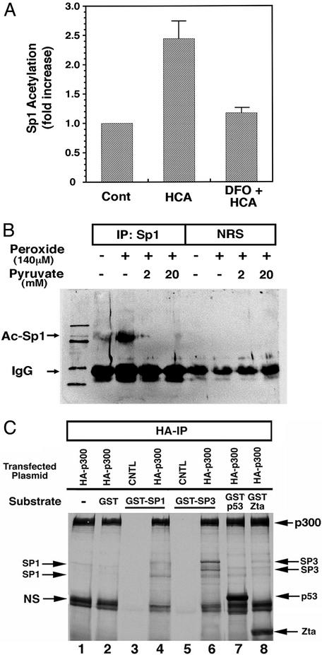
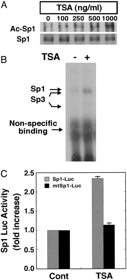
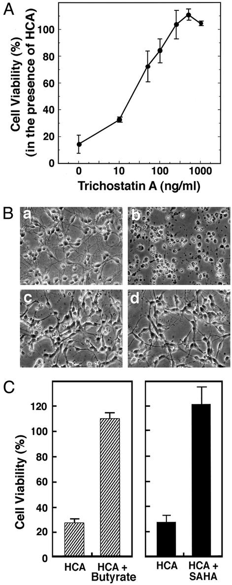
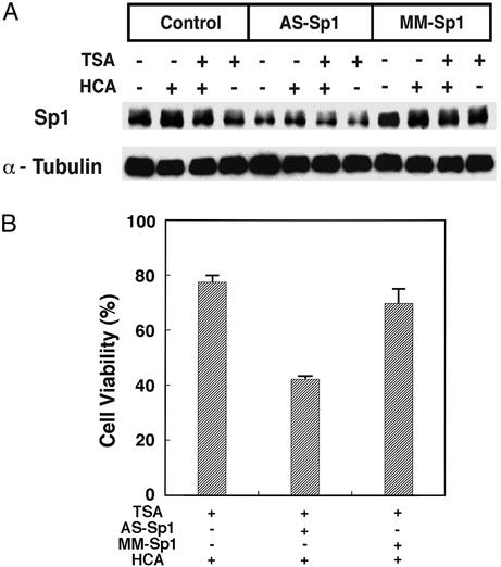
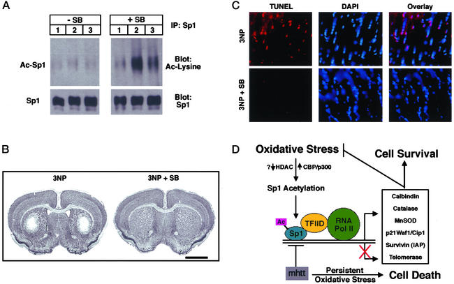
Oxidative stress is believed to be an important mediator of neurodegeneration. However, the transcriptional pathways induced in neurons by oxidative stress that activate protective gene responses have yet to be fully delineated. We report that the transcription factor Sp1 is acetylated in response to oxidative stress in neurons. Histone deacetylase (HDAC) inhibitors augment Sp1 acetylation, Sp1 DNA binding, and Sp1-dependent gene expression and confer resistance to oxidative stress-induced death in vitro and in vivo. Sp1 activation is necessary for the protective effects of HDAC inhibitors. Together, these results demonstrate that HDAC inhibitors inhibit oxidative death independent of polyglutamine expansions by activating an Sp1-dependent adaptive response.


Figures

Figure 1
Figure 1
Sp1 acetylation is increased by oxidative stress. (A) Immunoprecipitation of Sp1 followed by immunoblotting with an Ab to acetyl lysine residues reveals an increase in Sp1 acetylation in response to glutathione-depletion-induced oxidative stress. Coadministration of the antioxidant deferoxamine mesylate prevented Sp1 acetylation. Studies have established that deferoxamine (DFO) inhibits oxidative neuronal death distal to glutathione depletion (25). Results are mean fold increase of densitometric units ± SE for three separate experiments (P < 0.03). (B) U373 glioblastoma cell cultures were subjected to hydrogen peroxide with or without sodium pyruvate, harvested after 4 h, immunoprecipitated with anti-Sp1 Ab or normal rat serum (NRS), and analyzed by Western blots with anti-acetyl lysine Ab as described in Experimental Methods. (C) In vitro acetylation of recombinant Sp1 and Sp3 fusion proteins. Autoradiogram of SDS/PAGE gel of fractionated proteins subjected to the in vitro acetylation reaction. Arrows point to respective transcription factors containing radioactive acetyl groups in presence of the acetyl transferase hemagglutinin (HA)-p300. Differences in levels of acetylation by p300 among GST-Sp1, GST-Sp3, GST-p53, and GST-Zta principally reflect differing amounts of full-length protein loaded on gel as determined by Coomassie staining (data not shown).
Figure 2
Figure 2
HDAC inhibitors augment Sp1 acetylation, Sp1 DNA binding activity, and Sp1-dependent reporter gene expression in cortical neuronal cultures. (A) Sp1 acetylation levels in cortical neurons treated with the prototypic HDAC inhibitor TSA as determined by immunoprecipitation with an Sp1 Ab followed by immunoblotting with acetyl lysine Ab (Ac-Sp1) or Sp1 Ab alone (Sp1). Note that levels of Sp1 do not change with increasing concentrations of TSA. (B) TSA enhances binding of Sp1 and Sp3 to a canonical Sp1 DNA binding site. The electrophoretic mobility-shift assay was performed by using nuclear extracts from cortical neurons treated with and without TSA (100 ng/ml) for 60 min. The presence of Sp1 and Sp3 in each of the induced complexes was verified by supershift analysis. During this short period of TSA exposure, Sp1 or Sp3 protein levels did not change. (C) Sp1-dependent luciferase activity in control and TSA (100 ng/ml)-treated cortical neurons (gray bars). Note that luciferase activity does not change in the presence of TSA when the Sp1 response element has been mutated (black bars).
Figure 3
Figure 3
HDAC inhibitors inhibit neuronal death induced by glutathione-depletion-induced oxidative stress. (A) TSA inhibits HCA-induced apoptosis in a concentration-dependent manner. Cell viability was measured by 3-(4,5-dimethylthiazol-2-yl)-2,5-diphenyl tetrazolium bromide (MTT) reduction. Lactate dehydrogenase (LDH) release assay and TUNEL were performed in parallel to verify that MTT changes reflect changes in viability. Each point is the mean ± SD of three to five independent experiments. (B) Phase-contrast microscopy of cortical neurons: (a) control; (b) 1 mM HCA; (c) 100 ng/ml TSA (note the change in the morphology of the cell bodies); and (d) 100 ng/ml TSA plus 1 mM HCA (note how TSA-treated neurons maintain their cell body and neurite morphology in the presence of 1 mM HCA). (Magnifications: ×200.) (C) Structurally distinct HDAC inhibitors, butyrate (5 mM) and SAHA (5 μM), also inhibit HCA-induced death. Cell viability was measured by using the MTT reduction, LDH release, or TUNEL as described in Experimental Methods. All methods gave quantitatively similar results.
Figure 4
Figure 4
Sp1 is necessary for the protective effects of HDAC inhibitors. (A) Sp1 AS ODNs, but not MM ODNs, deplete Sp1 protein levels in cortical neurons. Sp1 ODNs do not alter the levels of α-tubulin. (B) Sp1 AS ODNs reverse TSA-induced prevention of HCA-induced death; MM ODNs do not. Results are mean ± SE for three separate experiments.
Figure 5
Figure 5
The HDAC inhibitor sodium butyrate enhances Sp1 acetylation and inhibits 3-NP-induced oxidative neuronal death in vivo. (A) Sp1 acetylation levels in brains of mice (n = 3 for each group) treated without (Left) and with (Right) the HDAC inhibitor sodium butyrate (SB;1.2 g/kg per day) along with 3-NP (50 mg i.p. twice a day for 5 days) as determined by immunoprecipitation with an Sp1 Ab followed by immunoblotting with acetyl lysine Ab (Ac-Sp1) or Sp1 Ab alone (Sp1). Note that levels of Sp1 do not change with SB treatment. (B) Nissl staining of a representative tissue section from mice treated as described in A. Note the loss of Nissl staining induced by 3-NP administration is completely inhibited by SB treatment. (C) TUNEL of a representative tissue section from mice treated as described in A. TUNEL labels double-stranded breaks in DNA and reflects DNA damage. DAPI (4′6-diamidino-2-phenylindole) intercalates into DNA and stains nuclei. Note overlay between TUNEL and DAPI in mice treated with 3-NP alone but not those treated with 3-NP plus SB. (Magnifications: ×400.) (D) Model for redox activation of Sp1 and adaptive responses to oxidative stress. Oxidative stress leads to enhanced acetylation of Sp1. Increased Sp1 acetylation could accrue from decreases in HDAC activity or increases in histone acetyl transferase activity. Hyperacetylated Sp1 binds DNA more avidly, leading to recruitment of coactivators such as TAFII130 in the TFIID complex. This putative Sp1-associated complex can then recruit RNA polymerase II to the promoter of genes such as catalase (22), MnSOD, and p21 waf1/cip1 and increase expression of their mRNA and protein. Up-regulation of the expression of these genes permits the cell to counter oxidative stress and oxidative damage and promotes cell survival. Mutant huntingtin protein can sequester Sp1 and TAFII130 and prevent appropriate adaptation to oxidative stress. The absence of these adaptive responses can lead to persistent oxidative stress and cell dysfunction and ultimately death.

References

    1. Sagara Y, Dargusch R, Chambers D, Davis J, Schubert D, Maher P. Free Radical Biol Med. 1998;24:1375–1389. - PubMed
    1. Atwood C S, Huang X, Moir R D, Tanzi R E, Bush A I. Met Ions Biol Syst. 1999;36:309–364. - PubMed
    1. Browne S E, Ferrante R J, Beal M F. Brain Pathol. 1999;9:147–163. - PMC - PubMed
    1. Castagne V, Gautschi M, Lefevre K, Posada A, Clarke P G. Prog Neurobiol. 1999;59:397–423. - PubMed
    1. Estevez A G, Crow J P, Sampson J B, Reiter C, Zhuang Y, Richardson G J, Tarpey M M, Barveito L, Beckman J S. Science. 1999;286:2498–2500. - PubMed

Publication types

MeSH terms