

Comparison of algorithms for detection of localised nerve fibre layer defects using scanning laser polarimetry
- PMID: 12642302
- PMCID: PMC1771596
- DOI: 10.1136/bjo.87.4.413
Comparison of algorithms for detection of localised nerve fibre layer defects using scanning laser polarimetry
Abstract
Aims: To evaluate different algorithms used to analyse retinal nerve fibre layer thickness (RNFL) data obtained by scanning laser polarimetry, in order to compare their relative abilities to discriminate between patients with glaucomatous localised nerve fibre layer defects and normal subjects.
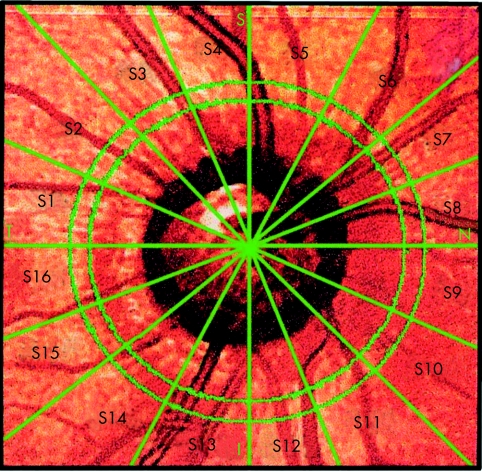
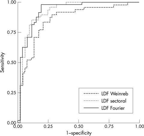
Methods: 48 eyes of 48 glaucomatous patients with localised RNFL defects and 53 eyes of 53 healthy subjects were included in this study. The localised RNFL defects were identified by RNFL photography and/or slit lamp biomicroscopic examination. All patients were submitted to RNFL examination using scanning laser polarimetry (GDx nerve fibre analyser, Laser Diagnostic Technologies, Inc, San Diego, CA, USA). Three methods of analysis of polarimetry data were used: GDx software provided parameters; RNFL thickness measurements in 16 equal sectors around the optic disc (sectoral analysis); and Fourier analysis of the curve of distribution of RNFL thickness measurements. Linear discriminant functions were developed to assess sensitivity and specificity of the sectoral based analysis and Fourier analysis and were compared to the GDx parameters. In addition, areas under the receiver operating characteristic (ROC) curves were compared.
Results: At a fixed specificity of 91%, the sensitivity of the linear discriminant function from sectoral data (LDF sectoral) was 81%, with an area under the ROC curve of 0.93. The linear discriminant function from Fourier measures had a comparable performance, with an area under the ROC curve of 0.93, and sensitivity of 71% for specificity at 91%. At the same specificity, the sensitivities of the GDx software provided parameters ranged from 15% to 40%. The areas under the ROC curves for the LDF sectoral and LDF Fourier were significantly greater than the ROC curve area for the single best GDx parameter.
Conclusion: The sectoral based analysis and the Fourier analysis of RNFL polarimetry data resulted in an improved detection of eyes with glaucomatous localised nerve fibre layer defects compared to the GDx software provided parameters.
Figures
References
-
- Sommer A, Katz J, Quigley HA, et al. Clinically detectable nerve fiber atrophy precedes the onset of glaucomatous field loss. Arch Ophthalmol 1991;109:77–83. - PubMed
-
- Tuulonen A, Lethola J, Airaksinen PJ. Nerve fiber layer defects with normal visual fields. Do normal optic disc and normal visual field indicate absence of glaucomatous abnormality? Ophthalmology 1993;100:587–98. - PubMed
-
- Jonas JB, Budde WM, Panda-Jonas S. Ophthalmoscopic evaluation of the optic nerve head. Surv Ophthalmology 1999;43:293–320. - PubMed
-
- Hoyt WF, Frisén LL, Newman NM. Fundoscopy of nerve fiber layer defects in glaucoma. Invest Ophthalmol Vis Sci 1973;12:814–29. - PubMed
Publication types
MeSH terms
LinkOut - more resources
Full Text Sources
Medical