

A photosensitive vascular smooth muscle store of nitric oxide in mouse aorta: no dependence on expression of endothelial nitric oxide synthase
- PMID: 12642395
- PMCID: PMC1573726
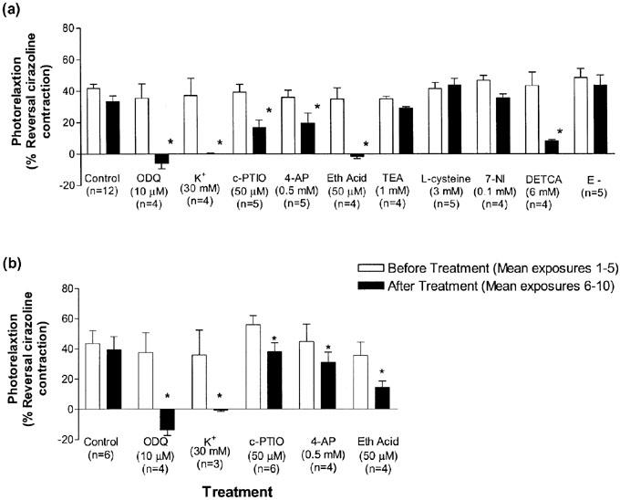
- DOI: 10.1038/sj.bjp.0705115
A photosensitive vascular smooth muscle store of nitric oxide in mouse aorta: no dependence on expression of endothelial nitric oxide synthase
Abstract
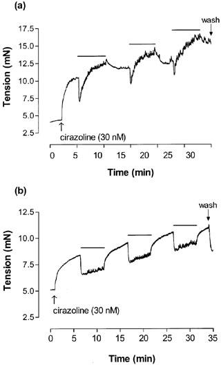
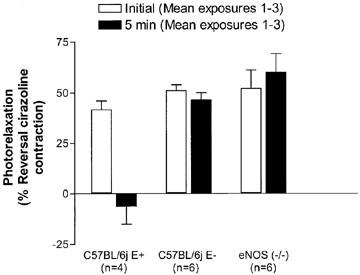
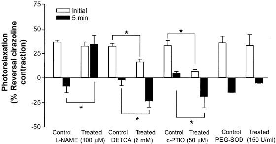
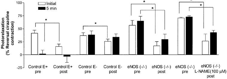
(1) Photorelaxation is the reversible relaxation of vascular smooth muscle (VSM) when irradiated with ultraviolet (UV) light resulting from the release of nitric oxide (NO). In this study we characterize the involvement of endothelial nitric oxide synthase (eNOS) in the photorelaxation response of thoracic aorta from endothelial NOS deficient (-/-) and control (C57BL/6j) mice. (2) Cirazoline contracted aortae were repeatedly exposed to 30 s of UV light every 3-4 min. Equal levels of photorelaxation (45+/-2%; n=34) was observed in both strains. (3) 1H-[1,2,4]-oxadiazolo[4,3-a]quinoxalin-1-one (ODQ), K(+), 2-(4-carboxyphenyl)-4,4,5,5-tetramethylimidazoline-1-oxyl-3-oxide (c-PTIO), 4-aminopyridine (4-AP) and ethacrynic acid significantly reduced the photorelaxation response. In C57BL/6j mice diethyldithiocarbamate (DETCA) also reduced photorelaxation. (4) Control endothelium-intact and -denuded aorta and L-NAME (100 micro M) treated and untreated eNOS (-/-) aortae were repeatedly exposed to UV light for 5 min every 10 min until no photorelaxation response was observed. After 1 h of rest in the dark the vessels showed between 30-70% recovery of the photorelaxation response indicating regeneration of the store in the absence of the endothelium and eNOS. (5) The results of this study suggest that photorelaxation in mouse aorta VSM results from the release of NO from a stable store of RSNOs, which activates soluble guanylate cyclase (sGC), leading to cGMP-dependent relaxation that is partially mediated by an increase in K(V) channel activation and hyperpolarization. In addition, the eNOS isoform is not essential for the formation of the photorelaxation store and a non-NOS source of NO may be involved in the maintenance of this store.
Figures

References
-
- AKAIKE T., YOSHIDA M., MIYAMOTO Y., SATO K., KOHNO M., SASAMOTO K., MIYAZAKI K., UEDA S., MAEDA H. Antagonistic action of imidazolineoxyl N-oxides against endothelium-derived relaxing factor (NO) through a radical reaction. Biochemistry. 1993;32:827–832. - PubMed
-
- ANDREWS K.L., MCGUIRE J.J., TRIGGLE C.R. Characterization of vascular smooth muscle photorelaxation in aorta from NOS knockout mice. The Pharmacologist. 2002;44:A214.
-
- ASKEW S.C., BUTLER A.R., FLITNEY F.W., KEMP G.D., MEGSON I.L. Chemical mechanisms underlying the vasodilator and platelet anti-aggregating properties of S-nitroso-N-acetyl-DL-penicillamine and S-nitrosoglutathione. Bioorg. Med. Chem. 1995;3:1–9. - PubMed
-
- BAUER J.A., FUNG H.L. Photochemical generation of nitric oxide from nitro-containing compounds: possible relation to vascular photorelaxation phenomena. Life Sci. 1994;54:L1–L4. - PubMed
Publication types
MeSH terms
Substances
LinkOut - more resources
Full Text Sources
Other Literature Sources
