Comparative Study
. 2003 Apr;121(4):277-86.
doi: 10.1085/jgp.200308791. Epub 2003 Mar 17.

The pore region of the skeletal muscle ryanodine receptor is a primary locus for excitation-contraction uncoupling in central core disease

Affiliations
Comparative Study

The pore region of the skeletal muscle ryanodine receptor is a primary locus for excitation-contraction uncoupling in central core disease

Guillermo Avila et al. J Gen Physiol. 2003 Apr.

Abstract

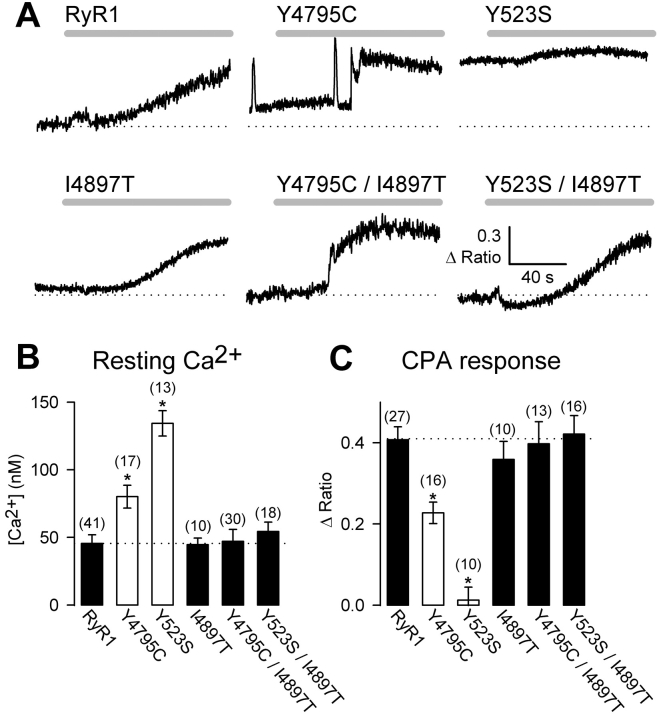
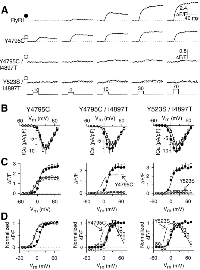
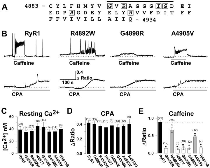
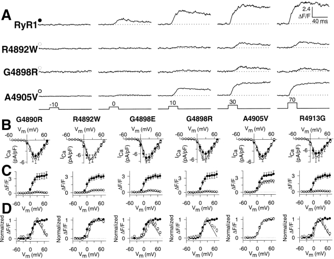
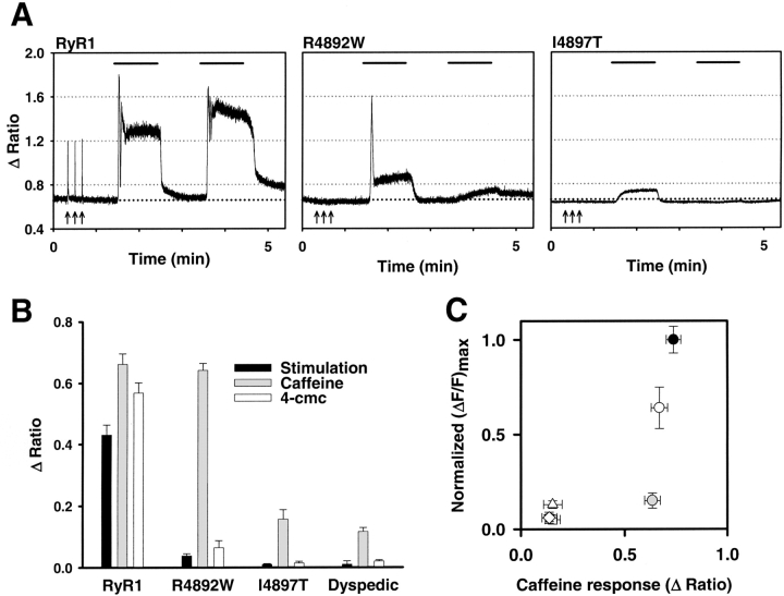
Human central core disease (CCD) is caused by mutations/deletions in the gene that encodes the skeletal muscle ryanodine receptor (RyR1). Previous studies have shown that CCD mutations in the NH2-terminal region of RyR1 lead to the formation of leaky SR Ca2+ release channels when expressed in myotubes derived from RyR1-knockout (dyspedic) mice, whereas a COOH-terminal mutant (I4897T) results in channels that are not leaky to Ca2+ but lack depolarization-induced Ca2+ release (termed excitation-contraction [EC] uncoupling). We show here that store depletion resulting from NH2-terminal (Y523S) and COOH-terminal (Y4795C) leaky CCD mutant release channels is eliminated after incorporation of the I4897T mutation into the channel (Y523S/I4897T and Y4795C/I4897T). In spite of normal SR Ca2+ content, myotubes expressing the double mutants lacked voltage-gated Ca2+ release and thus exhibited an EC uncoupling phenotype similar to that of I4897T-expressing myotubes. We also show that dyspedic myotubes expressing each of seven recently identified CCD mutations located in exon 102 of the RyR1 gene (G4890R, R4892W, I4897T, G4898E, G4898R, A4905V, R4913G) behave as EC-uncoupled release channels. Interestingly, voltage-gated Ca2+ release was nearly abolished (reduced approximately 90%) while caffeine-induced Ca2+ release was only marginally reduced in R4892W-expressing myotubes, indicating that this mutation preferentially disrupts voltage-sensor activation of release. These data demonstrate that CCD mutations in exon 102 disrupt release channel permeation to Ca2+ during EC coupling and that this region represents a primary molecular locus for EC uncoupling in CCD.


Figures

F<sc>igure</sc> 1.
Figure 1.
The I4897T CCD mutation blocks SR Ca2+ leak through Y523S and Y4795C release channels. (A) Representative CPA-induced Ca2+ responses (30 μM CPA, gray bars) in Indo-1 AM–loaded dyspedic myotubes expressing wild-type RyR1, Y523S, Y4795C, I4897T, Y523S/I4897T, Y4795C/I4897T. A dotted line representing the average resting fluorescence of RyR1-expressing myotubes is shown for comparison. (B) Average resting Ca2+ levels of dyspedic myotubes expressing wild-type RyR1, Y523S, Y4795C, I4897T, Y523S/I4897T, and Y4795C/I4897T. (C) Average steady-state CPA-induced Ca2+ responses (Δ Ratio = RCPA − Rbaseline) for dyspedic myotubes expressing wild-type RyR1, Y523S, Y4795C, I4897T, Y523S/I4897T, Y4795C/I4897T. Asterisks indicate significant differences (P < 0.05) compared with RyR1.
F<sc>igure</sc> 2.
Figure 2.
The I4897T mutation converts NH2-terminal (Y523S) and COOH-terminal (Y4795C) leaky SR Ca2+ release channels into EC uncoupled release channels. (A) Representative voltage-gated Ca2+ transients (ΔF/F) elicited by 30-ms test pulses to the indicated potentials in RyR1-, Y4795C-, Y4795C/I4897T-, and Y523S/I4897T-expressing dyspedic myotubes. Similar to that observed for Y523S-expressing myotubes (Avila and Dirksen, 2001), voltage-gated Ca2+ release activates at more negative potentials (i.e., −10 mV) in Y4795C-expressing dyspedic myotubes. (B) Average peak Ca2+ current activation curves for Y4795C- (left, open circles), Y4795C/I4897T- (middle, open circles), and Y523S/I4897T- (right, open circles) expressing dyspedic myotubes compared with that attributable to wild-type RyR1 (closed circles). (C) Average voltage dependence of peak intracellular Ca2+ transients (ΔF/F − V) for Y4795C- (left, open circles), Y4795C/I4897T- (middle, open circles), and Y523S/I4897T- (right, open circles) expressing dyspedic myotubes compared with that attributable to wild-type RyR1 (closed circles). The broken lines represent curves obtained from fitting data obtained for Y4785C- (middle) and Y523S- (right) expressing myotubes. (D) The ΔF/F − V data and curves in C are replotted following normalization to their respective maximal values. All data were acquired from three different sets of experiments and represent the average values from a total of 9 Y4795C-, 9 Y4795C/I4897T-, 8 Y523S/I4897T-, and 40 RyR1-expressing myotubes. The controls for each different set of experiments were plotted together with the corresponding experimental condition that was investigated simultaneously. The number of RyR1 controls collected for Y4795C-, Y4795C/I4897T-, and Y523S/I4897T-expressing myotubes were 18, 14, and 8, respectively.
F<sc>igure</sc> 3.
Figure 3.
The exon 102 CCD mutations in RyR1 exhibit normal resting Ca2+ levels and CPA-sensitive Ca2+ release, but a significant reduction in caffeine-induced Ca2+ release. (A) Amino acid sequence encoded by exon 102 of the human RyR1 gene. Seven different CCD mutations (G4891R, R4893W, I4898T, G4899E, G4899R, A4906V, and R4914G) have been identified for six different exon 102 amino acids (boxed letters), four of which are located within the putative pore-lining region of RyR1 (black bar). (B) Representative maximal caffeine- (10 mM, top) and CPA- (30 μM, bottom) induced Ca2+ responses in Indo-1 AM–loaded dyspedic myotubes expressing wild-type RyR1, R4892W, G4898R, and A4905V (numbers correspond to rabbit RyR1 sequence). The dotted line represents the average resting ratio of RyR1-expressing myotubes. (C–E) Average resting Ca2+ level (C), steady-state CPA response (D), and peak caffeine response (E) for dyspedic myotubes expressing wild-type RyR1 and each of the seven different exon 102 CCD mutations in RyR1. *, P < 0.01 compared with RyR1; π, P < 0.05 compared with RyR1.
F<sc>igure</sc> 4.
Figure 4.
Voltage-gated Ca2+ release is reduced by exon 102 CCD mutations in RyR1. (A) Representative voltage-gated Ca2+ transients (ΔF/F) elicited by 30-ms test pulses to the indicated potentials in RyR1-, R4892W-, G4898R-, and A4905V-expressing dyspedic myotubes. (B) Average peak Ca2+ current activation curves for G4890R-, R4892W, G4898E, G4898R, A4905V-, and R4913G-expressing myotubes (open circles) compared with that attributable to wild-type RyR1 (closed circles). (C) Average voltage dependence of peak intracellular Ca2+ transients for G4890R-, R4892W-, G4898E-, G4898R-, A4905V-, and R4913G-expressing myotubes (open circles) compared with that attributable to wild-type RyR1 (closed circles). (D) The ΔF/F − V data and curves in C normalized to their respective maximal values. Dyspedic myotubes expressing either the G4898E (n = 8), R4892W (n = 8), or the R4913G (n = 9) CCD mutants were compared with eight separate RyR1-expressing myotubes. Dyspedic myotubes expressing the G4890R (n = 8), A4905V (n = 10), or the G4898R (n = 8) CCD mutants were collected together with nine separate RyR1-expressing myotubes.
F<sc>igure</sc> 5.
Figure 5.
The R4892W mutation preferentially disrupts voltage-gated Ca2+ release. (A) Representative traces of electrical-, caffeine-, and 4-cmc–induced Ca2+ transients recorded from Indo-1 AM–loaded RyR1- (left), R4892W- (middle), and I4897T- (right) expressing myotubes. A brief train of supramaximal electrical stimuli (8 V for 20 ms at 0.1 Hz for 30 s) (arrows) was delivered before sequential applications of 10 mM caffeine (first bar), control Ringer's, 500 μM 4-cmc (second bar), and a final wash with control Ringer's. (B) Average electrically activated (black bars), caffeine-induced (gray bars), and 4-cmc–induced (white bars) Ca2+ transients for RyR1-, R4892W-, and I4897T-expressing myotubes. (C) The average magnitude of maximal voltage-gated Ca2+ release (obtained from experiments like those depicted in Fig. 4C) are plotted as a function of the average maximal caffeine response for RyR1 and each of the exon 102 CCD mutant RyR1 proteins. Symbols represent RyR1 (closed circle), G4890R (open diamond), R4892W (gray circle), G4898E (open triangle), G4898R (inverted open triangle), A4905V (open circle), and R4913G (open square). The symbols for the G4898R and R4913G mutants are plotted behind the symbol for G4890R.

References

    1. Avila, G., and R.T. Dirksen. 2000. Functional impact of the ryanodine receptor on the skeletal muscle L-type Ca2+ channel. J. Gen. Physiol. 115:467–480. - PMC - PubMed
    1. Avila, G., and R.T. Dirksen. 2001. Functional effects of central core disease mutations in the cytoplasmic region of the skeletal muscle ryanodine receptor. J. Gen. Physiol. 118:277–290. - PMC - PubMed
    1. Avila, G., J.J. O'Brien, and R.T. Dirksen. 2001. a. Excitation-contraction uncoupling by a human central core disease mutation in the ryanodine receptor. Proc. Natl. Acad. Sci. USA. 98:4215–4220. - PMC - PubMed
    1. Avila, G., K.M. O'Connell, L.A. Groom, and R.T. Dirksen. 2001. b. Ca2+ release through ryanodine receptors regulates skeletal muscle L-type Ca2+ channel expression. J. Biol. Chem. 276:17732–17738. - PubMed
    1. Dirksen, R.T. 2002. Bi-directional coupling between dihydropyridine receptors and ryanodine receptors. Front. Biosci. 7:d659–d670. - PubMed

Publication types

MeSH terms

Substances