. 2003 Mar 17;197(6):763-75.
doi: 10.1084/jem.20020150.

Detection of a functional hybrid receptor gammac/GM-CSFRbeta in human hematopoietic CD34+ cells

Affiliations

Detection of a functional hybrid receptor gammac/GM-CSFRbeta in human hematopoietic CD34+ cells

J Giron-Michel et al. J Exp Med. .

Abstract

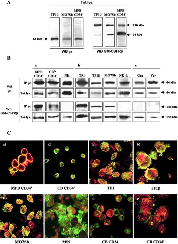
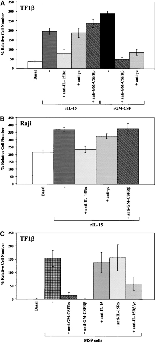
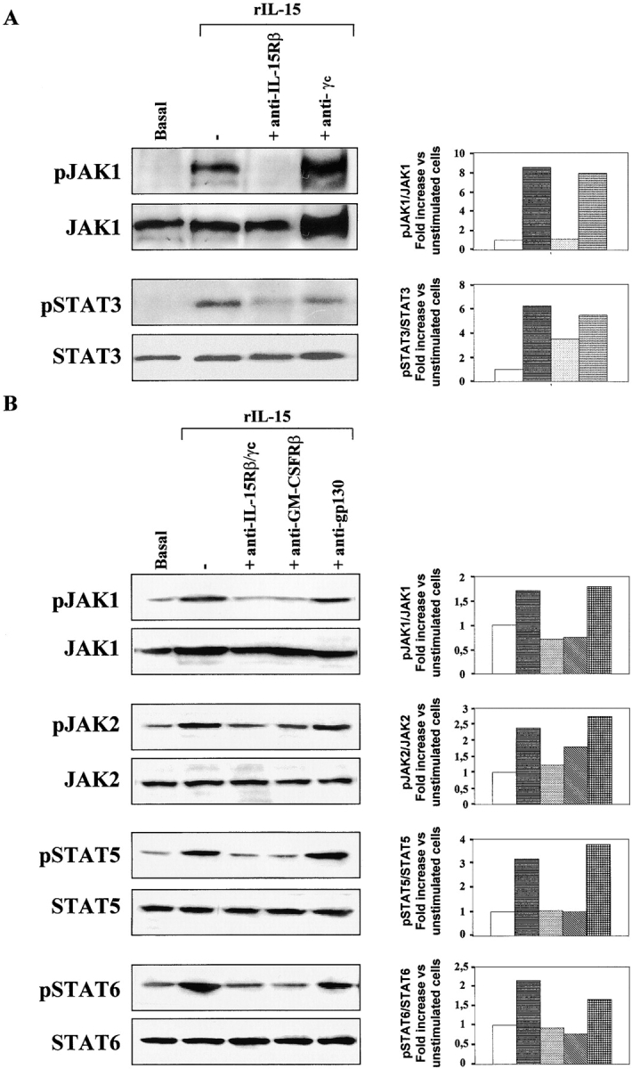
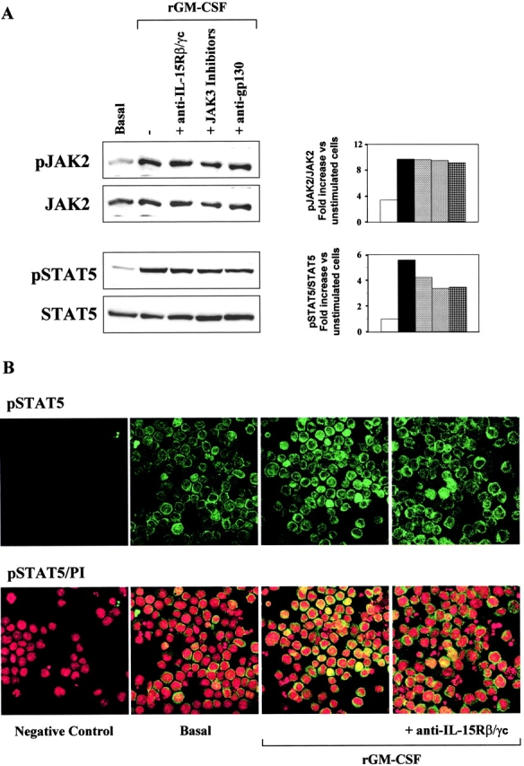
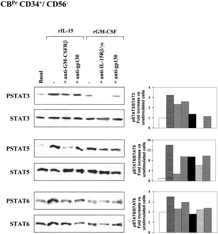
A functional hybrid receptor associating the common gamma chain (gammac) with the granulocyte/macrophage colony-stimulating factor receptor beta (GM-CSFRbeta) chain is found in mobilized human peripheral blood (MPB) CD34+ hematopoietic progenitors, SCF/Flt3-L primed cord blood (CB) precursors (CBPr CD34+/CD56-), and CD34+ myeloid cell lines, but not in normal natural killer (NK) cells, the cytolytic NK-L cell line or nonhematopoietic cells. We demonstrated, using CD34+ TF1beta cells, which express an interleukin (IL)-15Ralpha/beta/gammac receptor, that within the hybrid receptor, the GM-CSFRbeta chain inhibits the IL-15-triggered gammac/JAK3-specific signaling controlling TF1beta cell proliferation. However, the gammac chain is part of a functional GM-CSFR, activating GM-CSF-dependent STAT5 nuclear translocation and the proliferation of TF1beta cells. The hybrid receptor is functional in normal hematopoietic progenitors in which both subunits control STAT5 activation. Finally, the parental TF1 cell line, which lacks the IL-15Rbeta chain, nevertheless expresses both a functional hybrid receptor that controls JAK3 phosphorylation and a novel IL-15alpha/gammac/TRAF2 complex that triggers nuclear factor kappaB activation. The lineage-dependent distribution and function of these receptors suggest that they are involved in hematopoiesis because they modify transduction pathways that play a major role in the differentiation of hematopoietic progenitors.


Figures

Figure 1.
Figure 1.
γc/GM-CSFRβ interaction in human hematopoietic and nonhematopoietic cells. (A) Western blots of total lysates. MPB CD34+ cells, TF1, and M07Sb promyeloid CD34+ cell lines were analyzed for the expression for γc and GM-CSFRβ chains. Briefly, cell lysates were subjected to electrophoresis and the protein bands were transferred to PVDF membranes. The total lysate membranes (Tot.lys) were then probed with anti-γc or anti–GM-CSFRβ mAbs. The data shown are representative of three independent experiments. (B) Coimmunoprecipitation MPB CD34+ cells, CBPr CD34+/CD56 and normal polyclonal NK cells (panel a), TF1, TF1β, MO7sb promyeloid CD34+and NK-L cell lines (panel b) and human skin myofibroblasts (panel c) were analyzed for γc/GM-CSFRβ interactions. Briefly, total lysates were subjected to immunoprecipitation (IP) with an anti-γc antibody. Immunoprecipitates were subjected to electrophoresis and the protein bands were transferred to PVDF membranes. Membranes corresponding to the γc immunoprecipitate and to total lysates (Tot.lys) were probed with anti-γc or anti-GM-CSFRβ mAb. The data presented are representative of three independent experiments. (C) Confocal microscopy. Analysis by confocal microscopy of the interactions between the γc chain (green staining) and GM-CSFRβ (red staining) in MPB CD34+ cells (panel a1), CBPr CD34+ cells (panel a2), TF1 cells (panel b1), TF1β cells (panel b2), M07Sb (panel b3), and MS9 spleen myofibroblasts (panel c). As specificity controls, we analyzed the interactions between the γc chain (green staining) and IL-6R gp130 (red staining, panel d) or between the γc chain (green staining) and β1 integrin (red staining, panel e) in CB CD34+ cells. The images presented are compacted overlay pictures from serial optical sections, 1 μm thick, from the outside to the inner compartments of the cell. The yellow staining indicates colocalization of the various molecules. The data presented are representative of three independent experiments.
Figure 2.
Figure 2.
Proliferation induced by rIL-15 and rGM-CSF: IL-15R/GM-CSFR cross talk in TF1β cells. (A) Proliferative response of TF1β cells to recombinant IL-15 and GM-CSF. TF1β (IL-15Rα/β/γc) CD34+ cells were cultured for 4 d with rIL-15 or rGM-CSF (10 ng/ml) and their proliferation potential was then analyzed. Sister cultures were continuously incubated with neutralizing mAbs recognizing the IL-15Rα, γc, and GM-CSFRβ chains. In TF1β cells cultured with rIL-15, the proliferation rates of samples treated with an anti–GM-CSFRβ mAb were significantly higher (+25%; P < 0.001) than those of samples incubated with rIL-15 alone or with rIL-15 plus an anti-γc mAb. (B) Proliferative response of Raji cells to rIL-15. Raji cells (IL-15Rα/γc) were cultured for 4 d with rIL-15 (10 ng/ml) and analyzed for proliferation. Sister cultures were continuously incubated with neutralizing mAbs recognizing the IL-15Rα, γc, and GM-CSFRβ chains. (C) Proliferative response of TF1β cells to MS9 cells. TF1β cells were cocultured for 4 d with MS9 cells and analyzed for proliferation. Sister cultures were continuously incubated with neutralizing mAbs recognizing the GM-CSFRα, GM-CSFRβ, IL-15Rβ/γc, IL-15Rα chains, and IL-15. Cells were counted in an electronic Coulter counter and the data are expressed as % difference in proliferative potential with respect to control untreated samples. The data presented are representative of three independent experiments.
Figure 3.
Figure 3.
IL-15 signal transduction: IL-15R/GM-CSFR cross talk in TF1β cells. Analysis of JAK/STAT signal transduction by Western blotting. TF1β cells were incubated with 10 ng/ml rIL-15 for 15 min at 37°C. Sister cultures were pretreated for 1 h with neutralizing anti–IL-15Rβ/γc, anti-GM-CSFRβ, or anti-IL-6R gp130 mAbs. Cell extracts were analyzed by Western blotting using anti-phospho-JAK1 (pJAK1), anti-phospho-JAK2 (pJAK2), anti-phospho-TYK2 (pTYK2), anti-phospho-STAT5 (pSTAT5), and anti-phospho-STAT6 (pSTAT6) antibodies. Membranes were then reprobed with antibodies recognizing the native proteins. To correct for possible variations in the amount of protein loaded, values are expressed as pJAK/JAK or pSTAT/STAT ratios. pJAK/JAK and pSTAT/STAT levels were determined by densitometry including correction for background (NIH Image software). Results are expressed as increases (e.g., two times) with respect to untreated cells. The data presented are representative of three independent experiments.
Figure 4.
Figure 4.
GM-CSF signal transduction: IL-15R/GM-CSFR cross talk in TF1β cells. (A) Analysis of JAK2/STAT5 signal transduction by Western blotting. TF1β cells were incubated with 10 ng/ml rGM-CSF for 15 min at 37°C. Sister cultures were pretreated for 1 h with neutralizing anti–IL-15Rβ/γc mAbs or specific JAK3 inhibitors. Cell extracts were analyzed by Western blotting with anti-phospho-JAK2 (pJAK2) and anti-phospho-STAT5 (pSTAT5) antibodies. Membranes were then reprobed with antibodies recognizing the native proteins. To correct for possible variations in the amount of protein loaded, values are expressed as pJAK/JAK or pSTAT/STAT ratios. pJAK/JAK and pSTAT/STAT levels were determined by densitometry, including correction for background (NIH Image software). Results are expressed as an increase (e.g., two times) with respect to untreated cells. The data presented are representative of three independent experiments. (B) Analysis of pSTAT5 nuclear localization by confocal microscopy. TF1β cells were incubated with 10 ng/ml rGM-CSF for 15 min at 37°C. Sister cultures were pretreated for 1 h with neutralizing anti-IL-15Rβ/γc mAbs. Control (basal) and treated cultures were analyzed by confocal microscopy for pSTAT5 distribution in the cell, focusing particularly on whether this protein was present in the nucleus. Propidium iodide stains nuclei red whereas pSTAT5 is stained green. Yellow staining indicates the presence of pSTAT5 in the nucleus.
Figure 5.
Figure 5.
IL-15Rα/γc interaction in TF1 and TF1β cells. (A) Coimmunoprecipitation. TF1 and TF1β cells were analyzed for IL-15Rα/γc interactions. Briefly, lysates were subjected to immunoprecipitation (IP) with an anti-γc antibody. Immunoprecipitates were subjected to electrophoresis and the protein bands were transferred to PVDF membranes. Membranes corresponding to immunoprecipitated γc and total lysates (Tot.lys.) were probed with anti-γc or anti–IL-15Rα mAbs. The data presented are representative of three independent experiments. (B) Analysis of JAK3 phosphorylation by Western blotting. TF1 and TF1β cells were incubated with 10 ng/ml rIL-15 for 15 min at 37°C. Sister cultures were pretreated for 1 h with neutralizing anti–IL-15Rα, anti–GM-CSFRβ, and anti-gp130 mAbs. Cell extracts were subjected to immunoprecipitation with an anti-JAK3 mAb. JAK3 membranes were then re-probed with anti-phosphotyrosine 4G10 or anti-JAK3 mAbs. To correct for possible variations in the amount of protein loaded, values are expressed as pJAK3/JAK3 ratios. pJAK3/JAK3 levels were determined by densitometry, including correction for background (NIH Image software). Results are expressed as increases (e.g., two times) with respect to untreated cells. The data presented are representative of three independent experiments. (C) Coimmunoprecipitation. TF1 cells were analyzed for interactions between TRAF2, IL-15Rα, and γc. Briefly, lysates were subjected to immunoprecipitation (IP) with an anti-TRAF2 antibody. Immunoprecipitates were subjected to electrophoresis and the protein bands were transferred to PVDF membranes. Membranes corresponding to immunoprecipitated TRAF2 and total lysates (Tot.lys.) were probed with anti-TRAF2, anti-γc, or anti-IL-15Rα mAbs. The data presented are representative of three independent experiments. (D) Analysis of IκBα phosphorylation by Western blotting. TF1 and TF1β cells were incubated with 10 ng/ml rIL-15 or rIL-7 for 15 min at 37°C. Cell extracts were analyzed by Western blotting with anti-phospho-IκBα (pIκBα) antibody. Membranes were then reprobed with an antibody recognizing β-actin. To correct for possible variations in the amount of protein loaded, values are expressed as pIκBα/β actin ratios. pIκBα/β actin levels were determined by densitometry, including correction for background (NIH Image software). Results are expressed as increases with respect to untreated cells. The data presented are representative of three independent experiments.
Figure 6.
Figure 6.
IL-15 signal transduction: IL-15R/GM-CSFR cross talk in CBPr CD34+/CD56 cells. Analysis of STAT signal transduction by Western blotting. CB CD34+ cell populations were expanded by incubation for 5 d in the presence of SCF and Flt3-L. These cells displayed a CD34+/CD56 phenotype with no expression of lineage-specific markers. These CBPr CD34+/CD56 progenitors were incubated with 10 ng/ml rIL-15 or rGM-CSF for 15 min at 37°C. Sister cultures were pretreated for 1 h with neutralizing anti-IL-15Rβ/γc, anti-GM-CSFRβ, or anti-IL-6R gp130 mAbs. Cell extracts were analyzed by Western blotting with anti-phospho-STAT3 (pSTAT3), anti-phospho-STAT5 (pSTAT5), and anti-phospho-STAT6 (pSTAT6) antibodies. Membranes were then reprobed with antibodies recognizing the native proteins. To correct for possible variations in the amount of protein loaded, values are expressed as pSTAT/STAT ratios. pSTAT/STAT levels were determined by densitometry, including correction for background (NIH Image software). Results are expressed as increases with respect to untreated cells. The data presented are representative of two independent experiments.

Similar articles

Cited by

References

    1. Ratajczak, M.Z., and A.M. Gewirtz. 1995. The biology of hemopoietic stem cells. Semin. Oncol. 22:210–217. - PubMed
    1. Sharkis, S.J., C. Cremo, M.I. Collector, S.J. Noga, and A.D. Donnenberg. 1986. Thymic regulation of hematopoiesis: isolation of helper and suppressor populations using counterflow centrifugal elutriation. Blood. 68:787–789. - PubMed
    1. Majka, M., A. Janowska-Wieczorek, J. Ratajczak, K. Ehrenman, Z. Pietrzkowski, M.A. Kowalska, A.M. Gewirtz, S.G. Emerson, and M.Z. Ratajczak. 2001. Numerous growth factors, cytokines, and chemokines are secreted by human CD34(+) cells, myeloblasts, erythroblasts, and megakaryoblasts and regulate normal hematopoiesis in an autocrine/paracrine manner. Blood. 97:3075–3085. - PubMed
    1. Shibuya, A., K. Taguchi, H. Kojima, and T. Abe. 1991. Inhibitory effect of granulocyte-macrophage colony-stimulating factor therapy on the generation of natural killer cells. Blood. 78:3241–324. - PubMed
    1. Miller, J.S., F. Prosper, and V. McCullar. 1997. Natural killer (NK) cells are functionally abnormal and NK cell progenitors are diminished in granulocyte colony-stimulating factor-mobilized peripheral blood. Blood. 90:3098–3105. - PubMed

Publication types

MeSH terms

Substances