. 2003 Mar 17;197(6):777-88.
doi: 10.1084/jem.20021996.

Type-I interferon receptor deficiency reduces lupus-like disease in NZB mice

Affiliations

Type-I interferon receptor deficiency reduces lupus-like disease in NZB mice

Marie-Laure Santiago-Raber et al. J Exp Med. .

Abstract

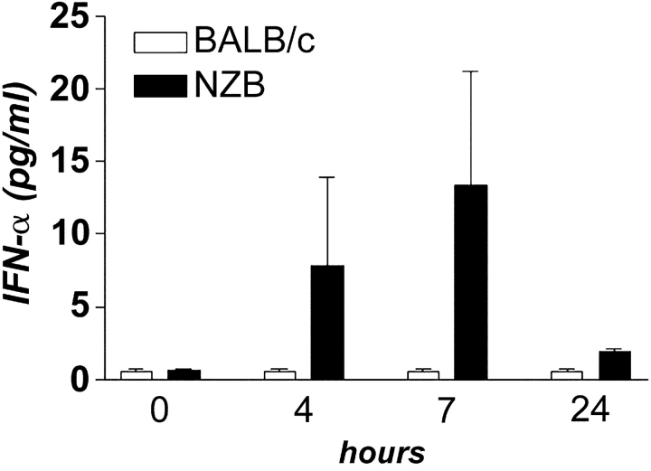
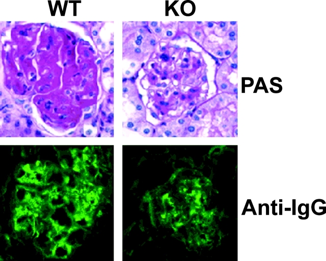
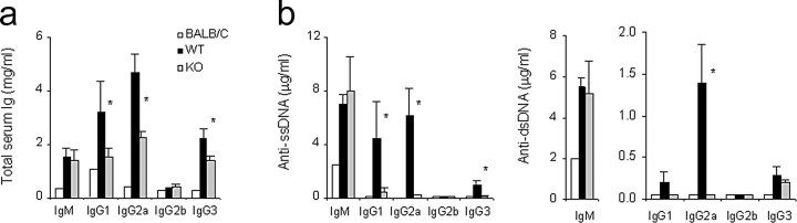
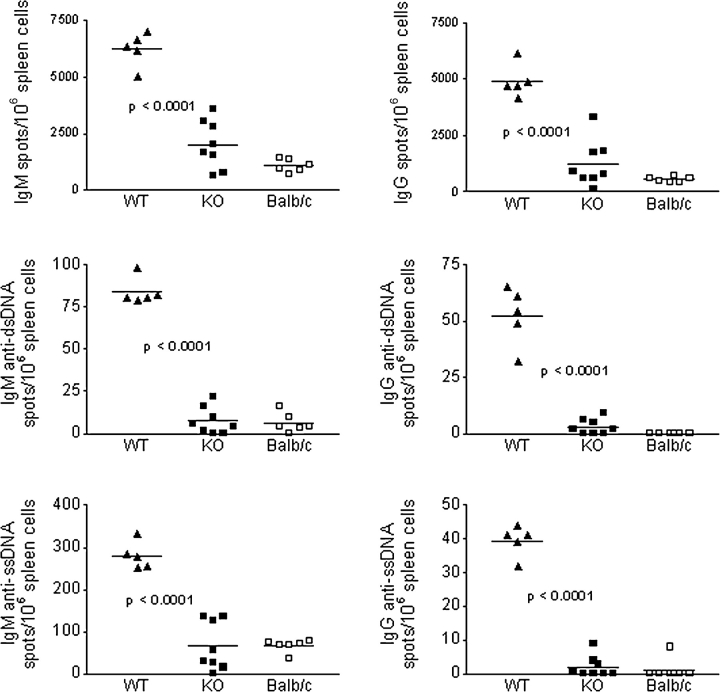
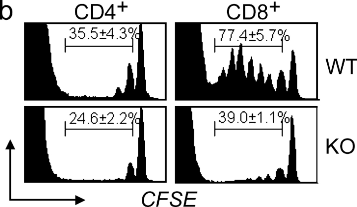
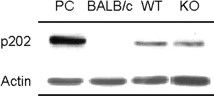
Indirect evidence suggests that type-I interferons (IFN-alpha/beta) play a significant role in the pathogenesis of lupus. To directly examine the contribution of these pleiotropic molecules, we created congenic NZB mice lacking the alpha-chain of IFN-alpha/betaR, the common receptor for the multiple IFN-alpha/beta species. Compared with littermate controls, homozygous IFN-alpha/betaR-deleted NZB mice had significantly reduced anti-erythrocyte autoantibodies, erythroblastosis, hemolytic anemia, anti-DNA autoantibodies, kidney disease, and mortality. These reductions were intermediate in the heterozygous-deleted mice. The disease-ameliorating effects were accompanied by reductions in splenomegaly and in several immune cell subsets, including B-1 cells, the major producers of anti-erythrocyte autoantibodies. Decreases of B and T cell proliferation in vitro and in vivo, and of dendritic cell maturation and T cell stimulatory activity in vitro were also detected. Absence of signaling through the IFN-alpha/betaR, however, did not affect increased basal levels of the IFN-responsive p202 phosphoprotein, encoded by a polymorphic variant of the Ifi202 gene associated with the Nba2 predisposing locus in NZB mice. The data indicate that type-I IFNs are important mediators in the pathogenesis of murine lupus, and that reducing their activity in the human counterpart may be beneficial.


Figures

Figure 1.
Figure 1.
Serum concentrations of IFN-α induced by poly (I:C). WT NZB (n = 3) and control BALB/c (n = 4) mice were bled before and, at different time points, after intraperitoneal injection of poly (I:C) and IFN-α levels were determined by ELISA.
Figure 2.
Figure 2.
Mortality rates and hemolytic anemia. (a) Cumulative survival of IFN-α/βR KO (n = 11), HT (n = 9), and WT (n = 9) NZB mice followed up to 12 mo; P = 0.015 for KO vs. WT; P > 0.05 for HT vs. WT. (b) Cumulative incidence of direct Coomb's test. Mouse groups were the same as in panel a. HT mice were examined at 12 mo only. (c) FACS® analysis of RBC-bound IgG autoantibodies in 10–12-mo-old mice. Each point represents the mean fluorescence intensity unit (FIU) from a single mouse and horizontal lines indicate the mean FIU; P = 0.004 for KO vs. WT; P = 0.04 for HT vs. WT mice. (d) Frequency of erythroblasts in spleens of 12-mo-old mice. RBC-depleted splenocyte suspensions were stained with an anti-erythroblast Ab (TER119) and analyzed by FACS®. Results of a representative mouse from each group are depicted. Numbers indicate percent of erythroblasts (mean ± SE) of 3–4 mice/group; P = 0.02 for KO or HT vs. WT mice; P = 0.05 for KO vs. HT.
Figure 3.
Figure 3.
Kidney immunohistology. Representative sections from 12-mo-old IFN-α/βR WT and KO NZB mice (n = 4 to 10) stained with PAS or FITC-conjugated anti–mouse IgG are shown.
Figure 4.
Figure 4.
Serum concentrations of (a) polyclonal Ig and (b) anti-ss and dsDNA autoantibodies. BALB/c mice (a pool of 3 individuals) and IFN-α/βR WT (n = 8), or KO (n = 7) NZB mice were analyzed at 10–12 mo. Bars indicate mean ± SE; *P < 0.05.
Figure 5.
Figure 5.
Frequency of splenic polyclonal Ig and anti-DNA AFCs. ELISPOT results for total IgM, total IgG, and corresponding anti-dsDNA or anti-ssDNA AFC in 12-mo-old BALB/c and IFN-α/βR WT or KO NZB mice are depicted. Data result from two separate experiments and each dot represents a single mouse. In all measurements, P < 0.0001 for WT vs. KO or BALB/c mice.
Figure 6.
Figure 6.
Frequency of peritoneal B-1 cells. Representative density plots of total peritoneal lymphocytes are shown, with numbers indicating percentages (mean ± SE) of gated B220+CD5+ cells in 12-mo-old IFN-α/βR WT and KO NZB mice (n = 3–6 mice/group); P = 0.01.
Figure 7.
Figure 7.
In vivo lymphocyte proliferation. (a) Spontaneous proliferation of splenic B and T cell subsets in 12 mo-old BALB/c, IFN-α/βR WT, and KO mice (n = 2–3/group) defined by BrdU-incorporation. Representative histograms and percentage of BrdUhi cells (mean ± SE) are shown. (b) Lymphopenia-induced homeostatic T cell proliferation. LN cells from 2–3 mo-old WT and KO mice were labeled with CFSE and infused into sublethally-irradiated WT NZB recipients (n = 3/group). Representative histograms at 8 d post-transfer and percentage (mean ± SE) of cells that had undergone 1 to 7 divisions are shown, with peaks of decreasing CFSE-intensity identifying sequential cellular divisions. Reduced in vivo proliferation of KO T cells was also observed 5 and 14 d after transfer (data not depicted).
Figure 7.
Figure 7.
In vivo lymphocyte proliferation. (a) Spontaneous proliferation of splenic B and T cell subsets in 12 mo-old BALB/c, IFN-α/βR WT, and KO mice (n = 2–3/group) defined by BrdU-incorporation. Representative histograms and percentage of BrdUhi cells (mean ± SE) are shown. (b) Lymphopenia-induced homeostatic T cell proliferation. LN cells from 2–3 mo-old WT and KO mice were labeled with CFSE and infused into sublethally-irradiated WT NZB recipients (n = 3/group). Representative histograms at 8 d post-transfer and percentage (mean ± SE) of cells that had undergone 1 to 7 divisions are shown, with peaks of decreasing CFSE-intensity identifying sequential cellular divisions. Reduced in vivo proliferation of KO T cells was also observed 5 and 14 d after transfer (data not depicted).
Figure 8.
Figure 8.
Maturation and function of DCs. (a) Effect of IFN-α on DC maturation. BM cells from IFN-α/βR WT and KO NZB mice were incubated with GM-CSF and IL-4 for 6 d, then with or without IFN-α for another 24 h to induce final maturation. The frequency of cells that had up-regulated MHC and B7 costimulatory molecules was determined by FACS® on gated live, CD11c+ cells. Basal levels (horizontal lines) for MHC class II, B7.1, and B7.2 were set on the basis of parallel stainings in the absence of the specific antibodies, and for MHC class I on the basis of constitutive expression defined with specific antibody. Percentages are the mean ± SE of three separate experiments with pooled cells from 3 mo-old mice (n = 2–4/pool); *P = 0.05. (b) DCs (3 × 10⁴) from IFN-α/βR WT, KO, and BALB/c mice, derived as described in panel a using either medium, IFN-α or LPS, were incubated with allogenic T cells (105) for 3 d and proliferative responses determined by H3-thymidine incorporation. Patterns of allostimulation remained similar when lower numbers (104) of DCs were used (data not depicted). Results are the mean ± SE of three separate experiments; *P < 0.05.
Figure 9.
Figure 9.
Expression of Ifi202-encoded p202. Protein extracts from spleens of BALB/c, IFN-α/βR WT, and KO NZB mice (n = 3) were analyzed by immunoblotting using an anti-p202 polyclonal antiserum. Representative results are shown. Expression levels (mean ± SE) of p202, calculated as percent of actin expression, were 13.6 ± 3.4% for WT mice, 12.2 ± 1.7% for KO mice, and 0.7 ± 0.2% for BALB/c mice. PC, positive control (recombinant p202).

Comment in

References

    1. Biron, C.A. 2001. Interferons alpha and beta as immune regulators–a new look. Immunity. 14:661–664. - PubMed
    1. Taniguchi, T., and A. Takaoka. 2001. A weak signal for strong responses: interferon-alpha/beta revisited. Nat. Rev. Mol. Cell Biol. 2:378–386. - PubMed
    1. Theofilopoulos, A.N., S. Koundouris, D.H. Kono, and B.R. Lawson. 2001. The role of IFN-gamma in systemic lupus erythematosus: a challenge to the Th1/Th2 paradigm in autoimmunity. Arthritis Res. 3:136–141. - PMC - PubMed
    1. Peng, S.L., J. Moslehi, and J. Craft. 1997. Roles of interferon-gamma and interleukin-4 in murine lupus. J. Clin. Invest. 99:1936–1946. - PMC - PubMed
    1. Balomenos, D., R. Rumold, and A.N. Theofilopoulos. 1998. Interferon-gamma is required for lupus-like disease and lymphoaccumulation in MRL-lpr mice. J. Clin. Invest. 101:364–371. - PMC - PubMed

Publication types

MeSH terms