

Review
doi: 10.1083/jcb.200212017.
Axonal transport of membranous and nonmembranous cargoes: a unified perspective
Affiliations
- PMID: 12642609
- PMCID: PMC2173776
- DOI: 10.1083/jcb.200212017
Item in Clipboard
Review
Axonal transport of membranous and nonmembranous cargoes: a unified perspective
J Cell Biol.
.
Abstract
Membranous and nonmembranous cargoes are transported along axons in the fast and slow components of axonal transport, respectively. Recent observations on the movement of cytoskeletal polymers in axons suggest that slow axonal transport is generated by fast motors and that the slow rate is due to rapid movements interrupted by prolonged pauses. This supports a unified perspective for fast and slow axonal transport based on rapid movements of diverse cargo structures that differ in the proportion of the time that they spend moving. A Flash feature (http://www.jcb.org/cgi/content/full/jcb.200212017/DC1) accompanies this Mini-Review.
Figures
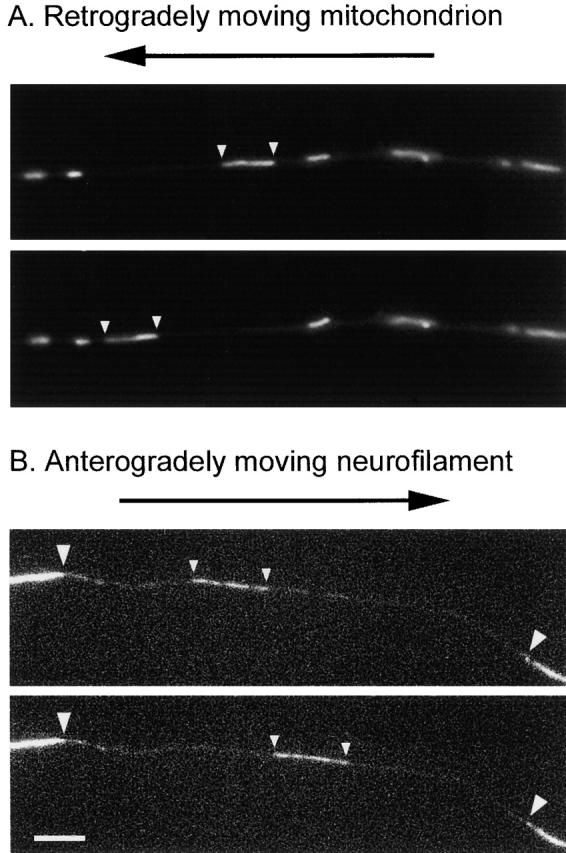
Movement of mitochondria and neurofilaments in axons. (A) Retrograde movement of a mitochondrion stained with Rhodamine-123 in the axon of a cultured neuron. The small arrowheads mark the position of the moving organelle. The other mitochondria in the field of view remain paused. The time interval between these two images is 3 s. (B) Anterograde movement of a GFP-tagged neurofilament in a photobleached axon of a cultured neuron. The large arrowheads mark the edges of the photobleached region, and the small arrowheads mark the leading and trailing ends of the filament. The time interval between these two images is 8 s. See Wang and Brown (2001) for details. Both neurofilaments and microtubules move in a rapid, intermittent, and bidirectional manner, which underscores the similarity in the underlying mechanism of movement of membranous and nonmembranous cargoes. Bar, 5 μm. Images in A were provided by Sunita R. Chada and Peter J. Hollenbeck. Images in B are reprinted from Molecular Biology of the Cell, 12:3257–3267, Wang, L., and A. Brown, Copyright 2001, with permission from American Society for Cell Biology.
References
-
- Almenar-Queralt, A., and L.S. Goldstein. 2001. Linkers, packages and pathways: new concepts in axonal transport. Curr. Opin. Neurobiol. 11:550–557. - PubMed
-
- Baas, P.W. 2002. Microtubule transport in the axon. Int. Rev. Cytol. 212:41–62. - PubMed
-
- Baas, P.W., and A. Brown. 1997. Slow axonal transport: the polymer transport model. Trends Cell Biol. 7:380–384. - PubMed
-
- Breuer, A.C., M.P. Lynn, M.B. Atkinson, S.M. Chou, A.J. Wilbourn, K.E. Marks, J.E. Culver, and E.J. Fleegler. 1987. Fast axonal transport in amyotrophic lateral sclerosis: an intra-axonal organelle traffic analysis. Neurology. 37:738–748. - PubMed
