. 2003 Apr 1;100(7):4078-83.
doi: 10.1073/pnas.0737556100. Epub 2003 Mar 17.

Mitochondrial pathology and apoptotic muscle degeneration in Drosophila parkin mutants

Affiliations

Mitochondrial pathology and apoptotic muscle degeneration in Drosophila parkin mutants

Jessica C Greene et al. Proc Natl Acad Sci U S A. .

Abstract

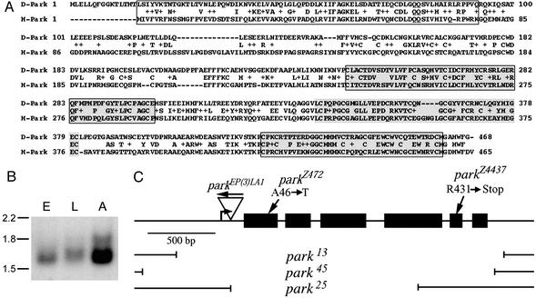
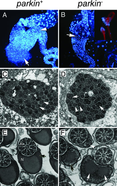
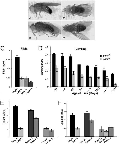
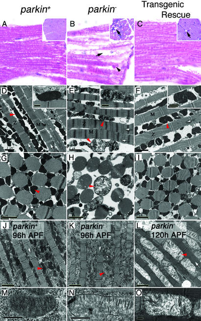
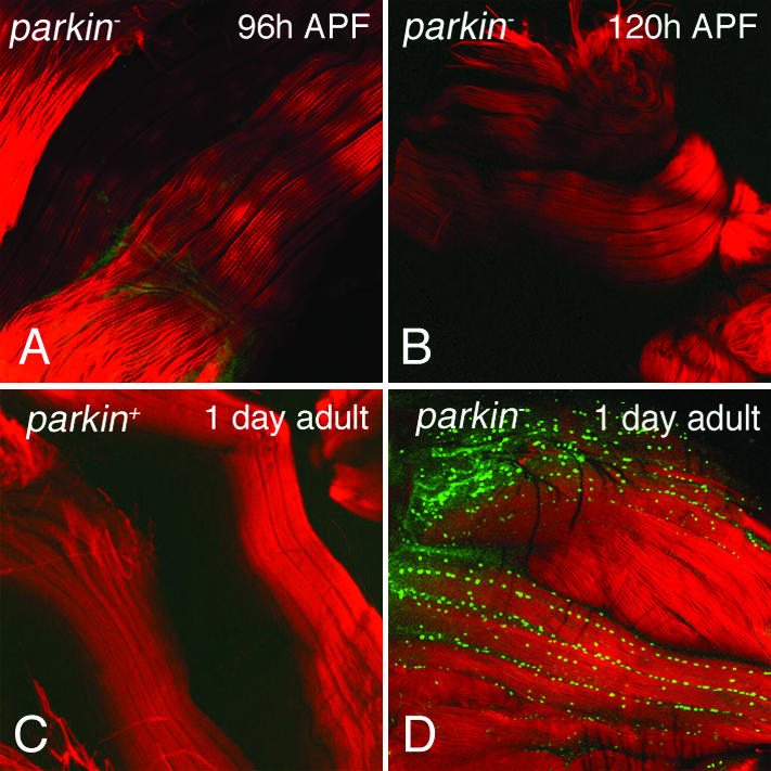
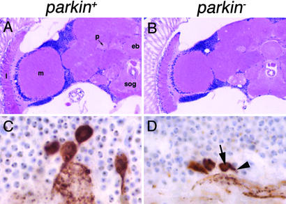
Parkinson's disease (PD) is a common neurodegenerative disorder characterized by loss of dopaminergic neurons in the substantia nigra. Several lines of evidence strongly implicate mitochondrial dysfunction as a major causative factor in PD, although the molecular mechanisms responsible for mitochondrial dysfunction are poorly understood. Recently, loss-of-function mutations in the parkin gene, which encodes a ubiquitin-protein ligase, were found to underlie a familial form of PD known as autosomal recessive juvenile parkinsonism (AR-JP). To gain insight into the molecular mechanism responsible for selective cell death in AR-JP, we have created a Drosophila model of this disorder. Drosophila parkin null mutants exhibit reduced lifespan, locomotor defects, and male sterility. The locomotor defects derive from apoptotic cell death of muscle subsets, whereas the male sterile phenotype derives from a spermatid individualization defect at a late stage of spermatogenesis. Mitochondrial pathology is the earliest manifestation of muscle degeneration and a prominent characteristic of individualizing spermatids in parkin mutants. These results indicate that the tissue-specific phenotypes observed in Drosophila parkin mutants result from mitochondrial dysfunction and raise the possibility that similar mitochondrial impairment triggers the selective cell loss observed in AR-JP.


Figures

Figure 1
Figure 1
Amino acid sequence, expression pattern, and mutant alleles of parkin. (A) Alignment of the amino acid sequences of Drosophila Parkin (D-Park) with human Parkin (H-Park). The N-terminal ubiquitin-like domain (boxed), RING finger domains (boxed and shaded), and In-Between Ring domain (shaded) are designated. (B) Northern blot analysis of poly(A)+ RNA obtained from wild-type embryos (E), third-instar larvae (L), and adults (A) using a parkin-specific probe. Size units are in kilobases. Adult flies appear to express a larger, less abundant parkin transcript in addition to the major transcript of 1.7 kb. (C) Molecular map of the parkin transcript showing the parkEP(3)LA1 insertion, the breakpoints of the three parkin deletion alleles described in this work, and the locations of the parkin point mutations. The bent arrow represents the predicted transcription initiation site of parkin and the black boxes designate Parkin protein-coding sequences. The arrow above the parkEP(3)LA1 insertion designates the orientation of this P element.
Figure 2
Figure 2
parkin mutants manifest a spermatid individualization defect associated with abnormal mitochondrial derivatives. (A) parkin+ testes reveal the presence of many individual sperm released from a ruptured testis (arrow). The long, thin nuclei of mature spermatozoa can also be seen inside the seminal vesicle (arrowhead). Round nuclei correspond to cells of the testis sheath. (B) Analysis of parkin testes reveals an absence of mature sperm in the seminal vesicle (arrow). However, mature spermatids are formed (Inset) but fail to individualize and remain as a syncytial bundle. (C) Ultrastructural analysis of a cross section through a wild-type late-stage 64-cell cyst showing the regular arrangement of developing spermatids (arrows). (E) Higher magnification cross sections of mature spermatozoa after individualization showing the axoneme (Ax) and a mitochondrial derivative, the Nebenkern (N), tightly enclosed in a membrane. Note the paracrystalline structure in the Nebenkern surrounded by uniform electron-dense material (arrow). In parkin mutants (D and F) the axonemes have a highly regular and well-formed appearance, but the Nebenkern display an abnormal distribution. Some spermatids are associated with multiple large Nebenkern (arrows), whereas others have a significantly diminished Nebenkern (arrowheads). In addition, the electron-dense matrix surrounding the paracrystalline structure of the Nebenkern appears diffuse (F, black arrow, compare with E). (A and B) Dissected testes were stained with DAPI to mark nuclei. (B Inset) Rhodamine-conjugated phalloidin was used to highlight spermatid tails. Genotypes; parkin+: parkrvA/Df(3L)Pc-MK, parkin: park13/Df(3L)Pc-MK.
Figure 3
Figure 3
Parkin function is required in mesoderm for normal wing posture, flight, and locomotion. (A and A′) Wing posture in 1-day-old control flies (parkrvA/Df(3L)Pc-MK) and (B and B′) age-matched parkin mutants (park25/Df(3L)Pc-MK) showing the downturned wing phenotype of parkin mutants. (C and D) parkin mutants exhibit impaired flight and climbing ability relative to control flies (see Methods for details). All flies tested were transheterozygous for the Df(3L)Pc-MK deletion and the parkin allele indicated. (E and F) Ectopic expression of parkin in mesoderm with the 24B-GAL4 (24) or Dmef2-GAL4 (21) driver restores flight and climbing ability in parkin mutants. All flies tested were transheterozygous for the Df(3L)Pc-MK deletion and the parkin allele indicated. Genotypes: Rescue 1: w; UAS-park/+; park13/24B-GAL4, Df; Rescue 2: w; UAS-park/+; park13/Dmef2-GAL4, Df; Control 1: w; park13/Dmef2-GAL4, Df; Control 2, w; park13/24B-GAL4, Df; Control 3: w; UAS-park/+; park13/Df. Error bars indicate the SEM.
Figure 4
Figure 4
Parkin mutants manifest muscle degeneration and mitochondrial pathology. (AC) Hematoxylin and eosin staining of longitudinal sections of IFMs show well preserved muscle in controls (A), contrasted with acute degeneration in parkin mutants (B) characterized by vacuole formation (arrow) and accumulation of cellular debris (arrowhead). Transgenic expression of parkin substantially restores muscle integrity (C), though occasional vacuoles are still seen (arrow). (Insets) Transverse section of IFMs. (D and G) Sections through parkin+ adult IFMs show a regular and compact myofibril arrangement (white arrows) with many electron-dense mitochondria (red arrowheads and Inset). (E and H) parkin adult IFMs show an irregular and dispersed myofibrillar arrangement with diffuse Z-lines and M-bands. Mitochondria are grossly swollen and malformed showing disintegration of cristae (red arrowheads and Inset). (F and I) Myofibril and mitochondrial integrity can be restored by transgenic expression of parkin in muscle tissue. (JO) The mitochondrial pathology is progressive and precedes myofibril degeneration. (J and M) IFMs from control 96-h pupae show many electron-dense mitochondria, whereas age-matched parkin mutants (K and N) already have mitochondria that are less electron-dense showing fewer cristae. (L and O) By 120 h, parkin pupae still show intact myofibril structure, but the mitochondria are profusely swollen as the cristae continue to degenerate. Genotypes: parkin+: w; parkrvA/Df(3L)Pc-MK, parkin: w; park25/Df(3L)Pc-MK, transgenic rescue: w; UAS-park; park25/24B-GAL4, park25. (Scale bars: DL, 2 μm; M–O and Insets in D–F, 0.5 μm.) Z, Z-lines; M, M-bands; APF, after puparium formation.
Figure 5
Figure 5
parkin mutants exhibit apoptotic cell death of flight muscle. IFMs from 96-h (A) and 120-h (B) parkin mutant pupae exhibit a lack of TUNEL-positive nuclei. One-day-old control flies (C) also lack TUNEL-positive nuclei, whereas age-matched parkin mutants (D) have many apoptotic nuclei (green). Phalloidin (red) highlights muscle tissue. Genotypes: parkin+: w; parkrvA/Df(3L)Pc-MK, parkin: w; park25/park25.
Figure 6
Figure 6
Loss of parkin function does not cause general neuronal degeneration or dopaminergic neuron loss. (A and B) Hematoxylin and eosin-stained frontal sections from control (A) compared with parkin mutant (B) flies at 30 days of age reveal appropriate organization of the nervous system including well-formed ellipsoid body (eb), peduncle (p), subesophageal ganglia (sog), medulla (m), and lamina (l). In addition, no age-related increase in neurodegeneration is evident in parkin mutants compared with controls in the cell cortex or neuropil. (C and D) Tyrosine hydroxylase immunostaining reveals similar number of neurons in the dorsomedial cluster in control flies (C) compared with parkin mutants (D), though shrinkage of the cell body (arrow) and decreased staining in the proximal dendrite (arrowhead) are frequently evident. Genotypes: parkin+: w; parkrvA/Df(3L)Pc-MK, parkin: w; park13/Df(3L)Pc-MK.

References

    1. Vaughan J R, Davis M B, Wood N W. Ann Hum Genet. 2001;65:111–126. - PubMed
    1. Gorell J, Johnson C, Rybicki B, Peterson E, Richardson R. Neurology. 1998;50:1346–1350. - PubMed
    1. Gwinn-Hardy K A. Movement Disorders. 2002;14:645–656. - PubMed
    1. Mizuno Y, Saitoh T, Sone N. Biochem Biophys Res Commun. 1987;143:294–299. - PubMed
    1. Nicklas W, Vyas I, Heikkila R. Life Sci. 1985;36:2503–2508. - PubMed

Publication types

MeSH terms